|
|
Post by jeany on Sept 25, 2009 8:58:56 GMT -5
Since we're studying the Nano aspect of our disease I thought this might be interesting for you. I've done a little research in the past at mdr on this Nanobacteria/Nanobe subject and found it very interesting. AND the new aspect that Nano is used in the production process of bio-insecticidal Baculovirus/Pheromones has also shown relevancy and might be another 'clue'. Here I'm showing how Nanobacteria leads to calcification..a form of calcium apatites in humans. This is the reason why I think that the cause respectively outbreak of our disease is related to a dysfunctional metabolism. www.pnas.org/content/95/14/8274.abstractNanobacteria: An alternative mechanism for pathogenic intra- and extracellular calcification and stone formation.
Calcium phosphate is deposited in many diseases, but formation mechanisms remain speculative. Nanobacteria are the smallest cell-walled bacteria, only recently discovered in human and cow blood and commercial cell culture serum. In this study, we identified with energy-dispersive x-ray microanalysis and chemical analysis that all growth phases of nanobacteria produce biogenic apatite on their cell envelope. Fourier transform IR spectroscopy revealed the mineral as carbonate apatite.The biomineralization in cell culture media resulted in biofilms and mineral aggregates closely resembling those found in tissue calcification and kidney stones. In nanobacteria-infected fibroblasts, electron microscopy revealed intra- and extracellular acicular crystal deposits, stainable with von Kossa staining and resembling calcospherules found in pathological calcification. Previous models for stone formation have led to an hypothesis that elevated pH due to urease and/or alkaline phosphatase activity is a lithogenic factor. Our results indicate that carbonate apatite can be formed without these factors at pH 7.4, at physiological phosphate and calcium concentrations. Nanobacteria can produce apatite in media mimicking tissue fluids and glomerular filtrate and provide a unique model for in vitro studies on calcification. Jeany |
|
|
|
Post by jeany on Sept 25, 2009 9:02:40 GMT -5
Here's another article about the pathogenic form of Nanobacteria:
Morphological, cultural, and immuno-histochemical characteristics of “Nanobacterium sanguineum” (NB) described in the literature are reviewed.
NB is reported to be a motile, Gram negative organism that divides by binary fission within a calcium-coated slimy shell; this yeast-like shell replicates by budding.
It measures between 20 and 200nm with a unique structure containing 16S ribosomal RNA.
NB has been observed by electron microscopy in coronary artery plaques (CAD) and in kidney stones (KS) found in renal diseases.
On the basis of supportive literature, we suggest that NB is not only present in the human body but also has auxiliary association with human ailments without a specific etiological role;
anti-NB antibody has been detected in subjects with calcified lesions and inflammation in diverse ailments including choriodecidual inflammation in pregnancy, ovarian cancers, arthritis and even Alzheimer’s disease.
More recent report on the detection and vertical transmission of NB antigen and anti-NB antibody in HIV-infected mothers supports the view that NB might be an important opportunistic infective agent contributing to HIV pathology;
we note that the presence of viable and transmitting NB was not studied and suggest further studies to establish vertical transmission of NB in HIV-infected persons.
On the basis of the foregoing we suggest that NB possibly exacerbates human ailments and raise the question:
Is NB a new life-form in search of human ailment or a commensal organism?
Recognizing the presence of NB in the human body, we discuss clinical trials, reported in the literature relevant to its eradication, with a rectal suppository containing very high amounts of disodium EDTA and tetracycline.
We suggest that tetracycline in this formulation acted in combination with EDTA, more as a chelating agent than an antibiotic; oxytetracycline- a non-chelating form of tetracycline-does not inhibit or kill NB.
Evaluation of anti-NB effect of orally administrable and potentially safer as well as therapeutically more acceptable chelating agent -ascorbic acid, acting alone or in combination with antibiotics-that eradicates another slime forming bacterium – Pseudomonas aeruginosa – in children with cystic fibrosis, is suggested.
Jeany
|
|
|
|
Post by jeany on Sept 25, 2009 9:16:27 GMT -5
Nanobes a possible nano-organismen.wikipedia.org/wiki/NanobeNanobes are tiny filamental structures first found in some rocks and sediments.  Some hypothesize that they are the smallest form of life, 1/10th the size of the smallest known bacteria. No conclusive evidence exists for whether these structures are, or are not, living organisms, and their classification is controversial. The smallest are just 20 nanometers in diameter. Some researchers believe that these structures are crystal growths, but the staining of these structures with dyes that bind to DNA might indicate that they are living organisms. Recently there has been some interest amongst bio-tech companies in commercial application of nanobes in utilization of plastics Nanobes are similar in size to nanobacteria, which are also structures that have been proposed to be extremely small living organisms. However, these two should not be confused. Nanobacteria are supposed to be cellular organisms, while nanobes are hypothesized to be a previously unknown form of life.It is a living organism (contains DNA or some analogue, and reproduces).Has a morphology similar to Actinomycetes and fungi.Nanobes are about 20 nm in diameter, which may be too small to contain the basic elements for an organism to exist (DNA, ribosomes, etc.), suggesting that if they grow and reproduce they would need to do so in an unconventional way. The Martian meteorite ALH84001, discovered in 1984 in the Antarctic, contained similar tubular structures which some astrobiologists suggested could be evidence of life at an earlier time on Mars. I'm not sure yet how this all fits together but, it could be a soil contaminator and reach the food source. If you look at the pictures it does look similar to Morg fibers/specimens. 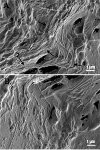 Ribbons and long filaments Ribbons and long filamentsRibbons of the web-structure as part of the microbial mat community, consisting mainly of long filaments with varying diameters. (A) Four ribbons can be distinguished within a small area (numbered arrows). The width of the ribbons is approximately 0.9 micrometer, their length cannot be defined: One ribbon appears from underneath at the lower edge of (B) and continues up to the rectangular "cross" of two filaments in (A), where it disappears in the mat. In these pictures most of the ribbons seem to follow the contour¡¦s of an underlying feature, probably a filament. (In all pictures the bar represents 1 micrometer, if not stated otherwise). 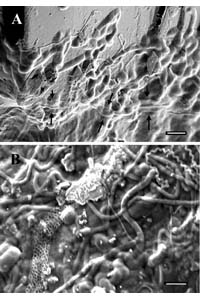 Web-structure integrated into the microbial mat community Web-structure integrated into the microbial mat community (A) Edge of a microbial mat attached to a mineral surface. The diameter of the smallest filaments is about 70 nm, the bigger ones of approximately 180 nm. Integrated in the mat is a ribbon starting at the left lower edge and continung horizontally (arrows). The web-structure is hardly recognizable because the meshes are filled with slipped-in material. Except for the middle part, the ribbon again seems to follow an underlying feature. (B) Mat community with clearly distinguishable microbes (rods 1.5 x 0.8 micrometer, cocci 1.1 x 0.7 micrometer, big filaments, spirillae and small filaments (diameter 300, 120 and 60 nm, resp.) and a ribbon (850 nm diameter) with a well-defined web-structure. In this case a "leading feature" underneath is not apparent. 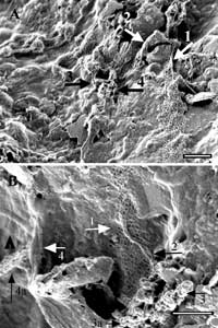
Variations of the shape of the ribbonsVariations of the shape of the ribbons: (A) The ribbon beginning with its normal appearance (lower right) is further up folding over a sharp ridge (arrow 1). After a 90 degree bend it curls on the upslope of a triangular pebble, and even more at the downslope, deflating to a compact strand with a diameter of 150 nm (arrow 2). Further to the left it resumes its normal structure. In the center (arrow 3), a part of the ribbon has totally collapsed; the remnants are partly discernible as a string of beads (arrow 4). (B) Twisting of the web (arrow 1, 2) leads to the formation of clusters (arrow 3), with bead-like components recognizable. The deflating area at left (4) arises from a folded (4a) and a “filled-up”ribbon (arrowhead). Bars = 1 micrometer. 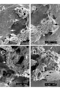
Twisting, folding and piling up as huge clustersThe ribbon on a smooth mineral surface (A, arrow) begins to fold up after a downward turn on a rougher surface, and curls up to two connected clusters, with remnants of the web-structure visible between these piles (arrowhead). Within the crumpling clusters (B = detail from A) hexagons of various size are recognizable (arrows), but even more pronounced in (C). Short chains and longer loop-forming strings are characteristic for the cluster in (D). 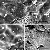
Bead-like substructures and the formation of loopsBead-like substructures and the formation of loops: (A) The bulk (center, right) consists of small strings (+/- 30 nm diameter) composed of tiny beads (better recognizable as individual rows or chains, arrows 1 = ~150 beads = 4.5 micrometer, 2 = ~84 beads = 2.5 micrometer). The strings show a tendency to form loops, frequently with an interspace close to that of the web-structures (arrows 3, 4, as examples); (B) beads at the upper rim, as part of the segments, within the interspace of a web, and very short strings; right foreground: clusters of nanobacteria (230 x 70 nm); (C) segments of an inflated ("puffed-up") web consisting of individual and lined-up beads (arrows); the beads also appear in the interspace of the ribbon structure. Jeany
|
|
|
|
Post by jeany on Sept 25, 2009 16:17:14 GMT -5
|
|
|
|
Post by jeany on Sept 25, 2009 16:20:00 GMT -5
Another aspect to consider: Nanobacteria in clouds could spread disease, scientists claim www.medicalnewstoday.com/articles/22413.phpThe radical theories about nanobacteria - micro-organisms considerably smaller than ordinary bacteria - in clouds are published in two recent articles in the Journal of Proteome Research by Dr Andrei P. Sommer of the University of Ulm, Germany, and Professor Chandra Wickramasinghe of Cardiff University, UK. They say nanobacteria are now accepted as being widely prevalent in the terrestrial environment and that their evidence is compelling for the existence of these nano-organisms, even in the stratosphere. In humans, nanobacteria have now been identified on four continents, they add. Experiments have shown that nanobacteria are excreted from the body in urine and their dispersal from the ground into the atmosphere and stratosphere appears to be inevitable. This happens because nanobacteria, lifted from the ground by winds, could transit between the high humidity region of the clouds and the relatively dry inter-cloud regions, leading to oscillations between a dormant state and one of activation," explained Professor Wickramasinghe.
"Remnants of a sticky protein (slime) coating nanobacteria makes them act as extremely efficient cloud condensation nuclei, with a tendency to aggregate to clusters upon contact." The contribution of nanobacteria to pathogenic bioaerosols, in the view of the authors, must overwhelm all other types of biological particles in the atmosphere. Jeany |
|
|
|
Post by jeany on Sept 25, 2009 16:24:04 GMT -5
Here's the connection to insects:www.pubmedcentral.nih.gov/articlerender.fcgi?artid=87844Infection with Bartonella weissii and Detection of Nanobacterium Antigens in a North Carolina Beef HerdVery recently, Bartonella organisms have been isolated from large ruminants (deer, elk, and dairy and beef cattle) located in the United States and in France. In this study, we report the serologic, microbiologic, and molecular findings related to the isolation of a Bartonella species in North Carolina beef cattle and the detection of nanobacterial antigen using a commercially available enzyme-linked immunosorbent assay. Bartonella spp. comprise an important group of vector-transmitted, intracellular bacterial pathogens. Diverse vectors, including sandflies, lice, mites, fleas, ticks, and potentially other insects can transmit these organisms among reservoir hosts. Although their actual existence, is controversial, nanobacteria are purportedly intracellular pathogens that can persist within intracellular compartments of the host for years. Nanobacteria appear to cause a variety of cytotoxic manifestations in cell culture, a feature that was responsible for their ultimate discovery. Based upon the respective 16S rRNA gene sequences, Bartonella and Nanobacterium spp. are members of the alpha subdivision of the Proteobacteria and purportedly share cross-reacting epitopes. In Europe, Kajander and colleagues have reported that more than 80% of commercial bovine serum lots contain Nanobacterium spp. . To our knowledge, data related to Nanobacterium infection in cattle is not yet available from the United States. As Nanobacteria purportedly share similar surface antigens with Bartonella spp., there may be serologic cross-reactivity; however, the extent to which cross-reactivity occurs has not been well characterized. Because of the potential for serologic cross-reactivity among these two organisms, we tested a subgroup of the herd with a commercially available antigen ELISA test kit. Surprisingly, 100% (22 of 22) of the cattle tested in this herd had very high levels of circulating nanobacterial antigens.
In humans and experimentally infected laboratory animals, Nanobacteria can cause chronic infection.Future studies should examine the potential relationship between exposure to and infection with Bartonella and Nanobacterium species, the extent to which these organisms might contribute to disease manifestations in cattle and the extent to which cattle might serve as a reservoir for human infection.
I'll look up what the symptoms of Bartonella are...btw..a co-infection to Lymes Disease... Jeany
|
|
|
|
Post by jeany on Sept 25, 2009 16:26:32 GMT -5
Bartonella en.wikipedia.org/wiki/BartonellaBartonella (formerly known as Rochalimaea) is a genus of Gram-negative bacteria. Facultative intracellular parasites, Bartonella species can infect healthy people but are considered especially important as opportunistic pathogens. Bartonella are transmitted by insect vectors such as ticks, fleas, sand flies and mosquitoes.  At least eight Bartonella species or subspecies are known to infect humans. In June 2007, a new species under the genus, called Bartonella melophagi, was discovered. This is the sixth species known to infect humans, and the ninth species and subspecies, overall, known to infect humans. In 2001 doctors treating Lyme disease first reported that their patients were co-infected with Bartonella. Multiple reports of this finding seem to indicate that Bartonella is not only a tick borne but a tick-transmitted pathogen. Treatment is dependent on which strain of Bartonella is found in a given patient. While Bartonella species are susceptible to a number of standard antibiotics in vitro—macrolides and tetracycline, for example—the efficacy of antibiotic treatment in immunocompetent individuals is uncertain. Immunocompromised patients should be treated with antibiotics because they are particularly susceptible to systemic disease and bacteremia. Drugs of particular effectiveness include trimethoprim-sulfamethoxazole, gentamicin, ciprofloxacin, and rifampin; B. henselae is generally resistant to penicillin, amoxicillin, and nafcillin Read also: en.wikipedia.org/wiki/Cat_scratch_feverJeany |
|
|
|
Post by jeany on Sept 25, 2009 16:28:41 GMT -5
Symptoms of Bartonella: Bartonella - Symptoms, Treatment and Prevention www.lymedisease.org/lyme101/coinfections/bartonella.htmlBartonellosis is often mild but in serious cases it can affect the whole body. Early signs are fever, fatigue, headache, poor appetite, and an unusual, streaked rash.  Swollen glands are typical, especially around the head, neck and arms. Burrascano suspects bartonellosis when neurologic symptoms are out of proportion to the other systemic symptoms of chronic Lyme. He also notes gastritis, lower abdominal pain, sore soles, and tender subcutaneous nodules along the extremities. Lymph nodes may be enlarged and the throat can be sore. Jeany |
|
|
|
Post by mfromcanada on Sept 25, 2009 21:16:03 GMT -5
Thanks Jeany, Great information and links
|
|
|
|
Post by jeany on Sept 26, 2009 5:10:51 GMT -5
Thanks Jeany, Great information and links Hi mfromcanada..you're welcome. It does look suspicious if you ask me and would explain why the people working on Morgs aren't able to find a similarity? and why our body can't rid it because our immune system doesn't recognize it as a foreign matter... Plus if you look at the insect aspect AND Bartonella a co-infection to Lymes..hmm.. Keeps me thinking.. Jeany.. btw..I was born in Edmonton, Alberta..hey! I'm Canadian too! nice to meet ya..how's the weather over there?. ;D |
|
|
|
Post by bannanny on Sept 26, 2009 18:22:10 GMT -5
Great info jeany... hard for me to follow, but nonetheless, great stuff! I was wondering about the dysfunctional metabolism and what it means. Since I got morgs, everything in my body has been up and down. First, I lost 20 lbs. in a very short time, then my blood pressure started going down slowly. It went all the way down to 90/50... they took it 3 times in the doctors ofc. once cuz they figured I should've been close to death it was so low! Then my cholesterol (which I've never ever had a problem with) started going up. Pretty soon, my blood pressure slowly started going back up too. It's now at 140/90 at it's highest, but normally it's running around 130/80. My pulse rate has always been high tho... usually runs around 102. So does all that have to do with a dysfunctional metabolism?
hugs ~~ bannanny
P.S. As always, your pics seem to talk to me!
|
|
|
|
Post by jeany on Oct 13, 2009 8:23:25 GMT -5
Shewanella putrefaciensen.wikipedia.org/wiki/Shewanella_putrefaciensShewanella putrefaciens is a Gram-negative marine bacterium. S. putrefaciens is also a facultative anaerobe with the ability to reduce iron and manganese metabolically; that is, it can use iron and manganese as the terminal electron acceptor in the electron transport chain (in contrast to obligate aerobes which must use oxygen for this purpose). It is also one of the organisms associated with the odour of rotting fish, as it is a marine organism which produces trimethylamines (hence the species name putrefaciens, from putrid). In both solid and liquid media, S. putrefaciens is often recognizable by its bright pink colour. On solid media, the colonies are round, fast-growing, and pink. The organism is also fast-growing in liquid media, and there will give the liquid an overall pink hue. Although it is very rare for it to act as a human pathogen, there have been cases of infections and bacteremia caused by S. putrefaciens.[1]  Biogenic formation of photoactive arsenic-sulfide nanotubes by Shewanellawww.pnas.org/content/104/51/20410.full Biogenic formation of photoactive arsenic-sulfide nanotubes by Shewanellawww.pnas.org/content/104/51/20410.fullHere, we report the production of an extensive extracellular network of filamentous, arsenic-sulfide (As-S) nanotubes (20–100 nm in diameter by ≈30 μm in length) by the dissimilatory metal-reducing bacterium Shewanella sp. HN-41. The As-S nanotubes, formed via the reduction of As(V) and S2O3 2−, were initially amorphous As2S3 but evolved with increasing incubation time toward polycrystalline phases of the chalcogenide minerals realgar (AsS) and duranusite (As4S). Upon maturation, the As-S nanotubes behaved as metals and semiconductors in terms of their electrical and photoconductive properties, respectively. The As-S nanotubes produced by Shewanella may provide useful materials for novel nano- and opto-electronic devices. The electrical characteristics of the As–S nanotubes were investigated by measuring the current–voltage (I–V) relationship. This suggested that the As–S filamentous nanotubes were functioning as nanowires. This result showed that the As–S filamentous nanotubes are photoactive. Jeany
|
|


