|
|
Post by jeany on Jul 26, 2009 11:01:57 GMT -5
What are Nanobacteria ? The term Nanobacteria is short for its scientific genus and species name Nanobacterium sanguineum, a Latin scientific term that means blood nanobacteria. Nanobacteria are nano-sized in that they are from 20-200 nanometers in size and are the smallest known self-replicating bacteria (a nanometer is 1 billionth of a meter and is approximately the width of ten hydrogen atoms side-to-side). Nanobacterium sanguineum is recognized as an emerging infectious disease. Chronic inflammatory responses are seen in areas where nanobacteria are found. Nanobacteria can infect any tissue or cell and have even been photographed actively killing T-6 Lymphocytes, an important component of our immune defense system. Nanobacteria are extremely small, slowly growing bacteria that can be cultured from the blood of humans and mammals. When compared to regular bacteria, these Nanobacteria are 1/100 to 1/1,000 the size, allowing them to easily move around into other cells and invade/infect them. Nanobacteria have the ability to kill human tissue cells, human immune system cells and other bacteria. Nanobacterial infection can cause alteration of cellular RNA and DNA gene-expression patterns in infected cells... this process can lead to genetic alteration, abnormal cell growth or proliferation rates. The nanobacteria-secreted calcium biofilm hardens around the nanobacteria forming a hard calcium apatite protective shell. In the calcified state, nanobacteria can either reproduce upon themselves forming aggregate budding-like clusters or they can just remain in a state of calcified dormancy. Our body does not recognize dormant calcified nanobacteria as a foreign substance or pathogen. When in calcified form, our bodies see nanobacteria as common calcium, a substance found throughout our bodies at all times. It is ONLY when they secrete their irritating endotoxic biofilm that our immune system becomes alarmed and responds with inflammation. Dormant Nanobacteria have the ability to be dormant and to become active at a later time. NanobacLabs now has available the nanobacTEST-S, the only blood test for Nanobacteria Antigen and Antibodies. The NanobacTEST-S checks for active nanobacterial infection and/or recent exposure to nanobacteria. NanobacLabs also has the NanobacTEST-U/A, a rapid urine screening test for nanobacteria that can be done in a doctor's office with results available in minutes. Nanobacteria have been isolated from IPV polio vaccine, Human Immune Gamma Globulin (IgG), Fetal Bovine Serum (FBS) and are therefore expected as potential contaminants in other human biologicals made with FBS. Nanobacteria are known to cause infections in humans, cattle, deer and are suspected to be infectious agents in other mammals. Chronic inflammatory responses are seen in areas where nanobacteria are found. Nanobacteria can infect any tissue or cell and have even been photographed actively killing T-6 Lymphocytes, an important component of our immune defense system. Nanobacteria has been shown by multiple scientific researchers to be the cause of pathological (disease-causing) calcification deposits in humans and mammals.If you have calcification deposits in your body that you were not born with, they are probably secondary to a nanobacterial infection. Some of the diseases involved with pathological calcification deposits are: Atherosclerotic Plaque, Coronary Artery Disease, Heart Plaque, Kidney Stones, Polycystic Kidney Disease (PKD), Cataracts, Glaucoma, CREST, Scleroderma, Psoriasis, Eczema, Lichen planus, www.aad.org/public/publications/pamphlets/common_lichen.htmlLiver Cysts, Breast Calcification, Prostate Calcification, Dental Plaque, Periodontal Disease, Dental Pulp Stones, Arthritis, Fibromyalgia, Brain Sand and some Cancers. Only nanobacteria cause tissue calcifications that grow at sub-saturation tissue levels.When explained to most physicians, their general response has been profound. www.hbci.com/~wenonah/history/rife.htmNanobacteria cause a "false positive" on Chlamydia ELISA testing. Since researchers have not cultured Chlamydia from atherosclerotic plaque after much effort, it seems probable that the positive Chlamydia ELISA tests were actually caused by Nanobacterium sanguineum. Most human biologicals and vaccines are made in fetal bovine serum, a medium that is known to be contaminated with nanobacteria. Also read how Vitamin C can help control Nanobacteria: How the Vitamins for preventing scurvy, beriberi, and pellagra were Discovered. Vitamin C in excess of our body's cellular needs provides the function of a chelating agent removing harmful materials such as lead from our bodies. You would need to consume about 20 grams of Vitamin C a day for a couple of weeks to realize any toxic symptoms. [ Citric Acid is another chelating agent that Nature provides for this purpose. So, keep a good amount of citrus fruits in your diet. Citrus also has bioflavonoids*, which are needed to help Vitamin C properly perform its functions. ] { * Bioflavonoids: a derivative of a flavone compound that helps maintain the capillary walls, reducing the likelihood of hemorrhaging......GRAPEFRUITSEEDEXTRACT!! Other studies that have reported supposedly harmful effects, have failed to realize that Vitamin C doesn't work alone in all cases. Bioflavonoids are required for Vitamin C to perform many of its functions and administering Vitamin C at high levels without providing its co–workers can account for the observations they made. Mercury has a greater affinity to Vitamin C that is present in the blood than it does for body tissue. Therefore, it provides a measure of protection against mercury poisoning. ....AMALGAM!!! And Salt: www.hbci.com/~wenonah/rid****/edta.htmUsing Disodium to disolve the shells of Nanobacteria EDTA as an Anionic Surfactant (Disodium Ethylene–Diamine–Tetra–Acetate Disodium inosinate - Wikipedia, the free encyclopedia Ethylenediaminetetraäcetic acid* ("EDTA") is a synthetic amino acid and was first used in the 1940's for treatment of heavy metal poisoning. It is widely recognized as effective for that use as well as certain others, including emergency treatment of hypercalcemia and the control of ventricular arrhythmias associated with digitalis toxicity. EDTA - Wikipedia, the free encyclopedia Its prominence as a chelating agent arises from its ability to "sequester" di- and tricationic metalions such as Ca2+ and Fe3+. After being bound by EDTA, metal ions remain in solution but exhibit diminished reactivity. Today, EDTA is mainly synthesised from ethylenediamine (1,2-diaminoethane), formaldehyde (methanal), and sodium cyanide.[2] This route yields the sodium salt, which can be converted in a subsequent step into the acid forms. Medicine EDTA is used to bond metal ions in chelation therapy, e.g. for mercury (performs poorly) and lead poisoning.[10] Similarly it is used to remove excess iron from the body. Dentists used EDTA solutions to remove inorganic debris (smear layer) and prepare root canals for obturation. In the laboratory, EDTA is widely used for scavenging metal ions: in biochemistry and molecular biology, ion depletion is commonly used to deactivate metal-dependent enzymes, either as an assay for their reactivity or to suppress damage to DNA or proteins. The fully dissolved EDTA is then usually mixed with Ascorbic Acid to adjust the solution pH to the proper level for use as an IV additive. Ascorbic Acid also has four OH– groups and will form stable metal salts, thus helping to bind "Free Radicals" and can provide a chelating function also. A properly mixed and diluted solution of EDTA and Vitamin C can also be given orally*. Nanobacteria.. biofilm producing....protectiv shell...feeding on heavy metals...Ascorbic acid and Salt....Detoxification. This all sounds familiar to me. Marc mentioned Biofilms with hardening shells on his site. Could be that this is the reason why the VitaminC/Salt protocol works against Morgellons..(for me it did) together with detox and immune system upbuilding. The symptoms of Nanobacterium sanguineum are similar to Morgellons Disease. NanobacLabs also has the NanobacTEST-U/A, a rapid urine screening test for nanobacteria that can be done in a doctor's office with results available in minutes. I think we should all do this test. Jeany |
|
|
|
Post by jeany on Jul 26, 2009 11:04:24 GMT -5
The Nanobacteria Revolution
The Nanobacteria Revolution :: Healthy Lifestyles and Natural Weight Loss Information | Fitness and Food Nutrition at Alive.com
Are heart disease, dental plaque, kidney stones, and a host of other calcium-deposit diseases really caused by an infection?
Research in the past decade suggests that diseases in which calcium accumulates inappropriately in the body (i.e., outside of your bones) may be caused by a bacterial infection.
Similarly, since 1990 there has been mounting evidence that abnormal calcification may be caused by a bacterial organism about 1/100 the size of a conventional bacterium, called Nanobacterium sanguineum (“nano” is the Latin word for very small or minute).
While other microbes like Chlamydia and assorted fungi have been implicated in the development of abnormal calcification, research indicates that the more likely cause may be nanobacteria.
Researchers E. Olavi Kajander and Neva Ciftcioglu discovered in 1995 that nanobacteria secrete a sticky, calcium-rich coating that allows them to adhere to cells inside artery walls and to each other.
The coating then calcifies into a shell, protecting the bacteria from the immune system as well as all antibiotics, radiation, and even chemotherapy.
An inflammatory cascade is initiated in the artery or organ that ultimately forms hard calcific plaque.
The plaque layers continually grow over a period of years, eventually leading to blood vessel or organ disease.
Due to their small size, nanobacteria slip through conventional filters and can contaminate vaccines and other biological treatments.
Studies discussed in The Calcium Bomb indicate that nanobacteria are found in human blood; they reproduce much more slowly than most viruses and bacteria; and they can be observed and cultured from the blood of healthy humans using special methods.
The idea that bacteria could be responsible for abnormal calcification in arteries, tissues, and organs is a very difficult concept to grasp in the context of a medical system that focuses on drugs and surgeries that treat the symptoms more often than the causes of disease. Most medical doctors believe that the cause of abnormal calcification is, as yet, unexplained.
Dark field microscopy and various blood culturing techniques have demonstrated the presence of numerous bacteria and fungi circulating in the bloodstreams of healthy people, including Bartonella, Brucella, and Candida.
A study published in the Journal of Clinical Microbiology (March 2001) shows the presence of nanobacteria in beef cattle.
Human infection with nanobacteria may be the result of consuming meat from infected cows and getting contaminated bovine serum vaccines.
If you have ever had a vaccine, you may harbour nanobacteria in your blood, tissues, and organs.
There are basically two ways to determine if nanobacteria are present in your body.
Naturopathic doctors in Canada can run tests for nanobacteria by sending patients’ blood samples to a lab in Montreal.
The Calcium Bomb also contains information on how to order home kits to test for nanobacteria.
The most accurate test is a blood test that measures both antigens and antibodies linked to nanobacteria.
Another method is a special type of cardiac CT scan that calculates something called a calcium score. The higher the calcium score, the more likely the individual suffers from a nanobacterial infection.
A recent variation of standard intravenous chelation therapy currently works best to reverse diseases caused by nanobacteria.
EDTA (ethylenediamine tetra-acetic acid) chelation therapy uses an amino acid solution to dissolve calcium deposits in the body.
There it is: amino acids!!...L-Arginine..NAC!!
The way to kill nanobacteria and reverse the diseases they cause is to strip away their calcium shell with EDTA and then attack the suddenly exposed nanobacteria with tetracycline.
To date, this is the only antibiotic proven to be effective.
It is conceivable that natural alternatives to prescription antibiotics could be used (e.g., oil of oregano, berberine, colloidal silver, etc.)
Dr. Roberts suggests that a combination of oral EDTA with supportive antioxidants plus an EDTA rectal suppository and 500 mg of tetracycline all given once daily before bedtime is the most effective way of ridding the body of both the pathological calcium deposits and the nanobacteria.
Jeany
|
|
|
|
Post by jeany on Jul 26, 2009 11:05:50 GMT -5
Facts About Tetracycline Antibiotics
Tetracycline antibiotics are broad-spectrum bacteriostatic agents, that inhibit bacterial protein synthesis and may be effective against a wide variety of microorganisms.
These medications are used in the treatment of infections of the respiratory tract, sinuses, middle ear, urinary tract, skin, intestines. They are also used to treat Gonorrhoea. Their most common current use is in the treatment of moderately severe acne and rosacea.
The most commonly prescribed tetracycline antibiotics are: tetracycline, doxycycline, minocycline, oxytetracycline.
Jeany
|
|
|
|
Post by jeany on Jul 26, 2009 11:06:54 GMT -5
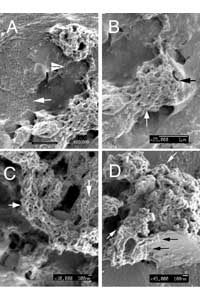
Nanobacteria: An alternative mechanism for pathogenic intra- and extracellular calcification and stone formation
Nanobacteria: An alternative mechanism for pathogenic intra- and extracellular calcification and stone formation ? PNAS
Calcium phosphate is deposited in many diseases, but formation mechanisms remain speculative.
Nanobacteria are the smallest cell-walled bacteria, only recently discovered in human and cow blood and commercial cell culture serum.
In this study, we identified with energy-dispersive x-ray microanalysis and chemical analysis that all growth phases of nanobacteria produce biogenic apatite on their cell envelope.
Fourier transform IR spectroscopy revealed the mineral as carbonate apatite.
The biomineralization in cell culture media resulted in biofilms and mineral aggregates closely resembling those found in tissue calcification and kidney stones.
In nanobacteria-infected fibroblasts, electron microscopy revealed intra- and extracellular acicular crystal deposits, stainable with von Kossa staining and resembling calcospherules found in pathological calcification.
Previous models for stone formation have led to an hypothesis that elevated pH due to urease and/or alkaline phosphatase activity is a lithogenic factor.
Our results indicate that carbonate apatite can be formed without these factors at pH 7.4, at physiological phosphate and calcium concentrations.
Nanobacteria can produce apatite in media mimicking tissue fluids and glomerular filtrate and provide a unique model for in vitro studies on calcification.
Ok...let me see what biogenic apatite means...
Jeany
|
|
|
|
Post by jeany on Jul 26, 2009 11:09:15 GMT -5
biogenic [bye-oh-jen-ik] originating from a living organism Apatite Apatite - Wikipedia, the free encyclopedia Apatite is a group of phosphate minerals. Apatite is one of few minerals that are produced and used by biological micro-environmental systems. Apatite has a Mohs Scale hardness of 5. Hydroxylapatite is the major component of tooth enamel. Similarly, fluoridated water allows exchange in the teeth of fluoride ions for hydroxyl groups in apatite. Too much fluoride results in dental fluorosis and/or skeletal fluorosis. In the United States, apatite is often used to fertilize tobacco. The primary use of apatite is in the manufacture of fertilizer - it is a source of phosphorus. Fluoro-Chloro Apatite forms the basis of the, now obsolete, Halophosphor fluorescent tube phosphor system. 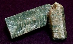  Jeany |
|
|
|
Post by jeany on Jul 26, 2009 11:10:17 GMT -5
The Crystal Structure of Apatite The Crystal Structure of Apatite, Ca5(PO4)3(F,OH,Cl) -- Hughes and Rakovan 48 (1): 1 -- Reviews in Mineralogy and Geochemistry it is the major source of phosphorous, both as an ore and the base of the global phosphorous cycle. As the major ore mineral of phosphorous, apatite is critical for the production of huge quantities of fertilizers, detergents and phosphoric acid; the extracted phosphorous is also used in many other applications such as phosphors, rust removers, motor fuels, and insecticides to name but a few.  Jeany |
|
|
|
Post by jeany on Jul 26, 2009 11:11:09 GMT -5
Resume:
Nanobacteria found in human and cow blood samples....produce a sticky biofilm...mimics tissue fluids....forms crystal like structures 'apatites'..
apatites... different colors such as blue, green, yellow and red...a form of phosphorus...found in soil...used as a fertilizer...insecticides..used also a remediation for contaminated soils...motor fuels...
and this is polluting our WATER!!
Another thought: what is, if this interacts with Fluoride...? in water...toothpaste?
Jeany
|
|
|
|
Post by jeany on Jul 26, 2009 11:12:06 GMT -5
Here's another article about the pathogenic form of Nanobacteria:
Elsevier: Article Locator
Morphological, cultural, and immuno-histochemical characteristics of “Nanobacterium sanguineum” (NB) described in the literature are reviewed.
NB is reported to be a motile, Gram negative organism that divides by binary fission within a calcium-coated slimy shell; this yeast-like shell replicates by budding.
It measures between 20 and 200nm with a unique structure containing 16S ribosomal RNA.
NB has been observed by electron microscopy in coronary artery plaques (CAD) and in kidney stones (KS) found in renal diseases.
On the basis of supportive literature, we suggest that NB is not only present in the human body but also has auxiliary association with human ailments without a specific etiological role; anti-NB antibody has been detected in subjects with calcified lesions and inflammation in diverse ailments including choriodecidual inflammation in pregnancy, ovarian cancers, arthritis and even Alzheimer’s disease.
More recent report on the detection and vertical transmission of NB antigen and anti-NB antibody in HIV-infected mothers supports the view that NB might be an important opportunistic infective agent contributing to HIV pathology;
we note that the presence of viable and transmitting NB was not studied and suggest further studies to establish vertical transmission of NB in HIV-infected persons.
On the basis of the foregoing we suggest that NB possibly exacerbates human ailments and raise the question:
Is NB a new life-form in search of human ailment or a commensal organism?
Recognizing the presence of NB in the human body, we discuss clinical trials, reported in the literature relevant to its eradication, with a rectal suppository containing very high amounts of disodium EDTA and tetracycline.
We suggest that tetracycline in this formulation acted in combination with EDTA, more as a chelating agent than an antibiotic; oxytetracycline- a non-chelating form of tetracycline-does not inhibit or kill NB.
Evaluation of anti-NB effect of orally administrable and potentially safer as well as therapeutically more acceptable chelating agent -ascorbic acid, acting alone or in combination with antibiotics-that eradicates another slime forming bacterium – Pseudomonas aeruginosa – in children with cystic fibrosis, is suggested.
soo...antibiotics...chelation (detoxification) and Vitamin C eradicates slime producing bacterium? hmm...very interesting!!
NAC is a 'detoxer' together with Vitamin C (ascorbin acid) and antibiotics such a Tetracycline KILLS Nanobacteria!!
There ya go!
Jeany
|
|
|
|
Post by jeany on Jul 26, 2009 11:14:17 GMT -5
Nanobes a possible nano-organism Nanobe - Wikipedia, the free encyclopedia Nanobes are tiny filamental structures first found in some rocks and sediments.  Some hypothesize that they are the smallest form of life, 1/10th the size of the smallest known bacteria. No conclusive evidence exists for whether these structures are, or are not, living organisms, and their classification is controversial. The smallest are just 20 nanometers in diameter. Some researchers believe that these structures are crystal growths, but the staining of these structures with dyes that bind to DNA might indicate that they are living organisms. Recently there has been some interest amongst bio-tech companies in commercial application of nanobes in utilization of plastics Nanobes are similar in size to nanobacteria, which are also structures that have been proposed to be extremely small living organisms. However, these two should not be confused. Nanobacteria are supposed to be cellular organisms, while nanobes are hypothesized to be a previously unknown form of life. * It is a living organism (contains DNA or some analogue, and reproduces). * Has a morphology similar to Actinomycetes and fungi. * Nanobes are about 20 nm in diameter, which may be too small to contain the basic elements for an organism to exist (DNA, ribosomes, etc.), suggesting that if they grow and reproduce they would need to do so in an unconventional way. * The Martian meteorite ALH84001, discovered in 1984 in the Antarctic, contained similar tubular structures which some astrobiologists suggested could be evidence of life at an earlier time on Mars. Jeany |
|
|
|
Post by jeany on Jul 26, 2009 11:17:32 GMT -5
Nanoben#2: figuren Ribbons and long filaments 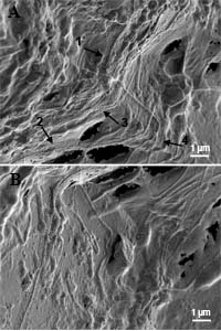 Ribbons of the web-structure as part of the microbial mat community, consisting mainly of long filaments with varying diameters. (A) Four ribbons can be distinguished within a small area (numbered arrows). The width of the ribbons is approximately 0.9 micrometer, their length cannot be defined: One ribbon appears from underneath at the lower edge of (B) and continues up to the rectangular "cross" of two filaments in (A), where it disappears in the mat. In these pictures most of the ribbons seem to follow the contour¡¦s of an underlying feature, probably a filament. (In all pictures the bar represents 1 micrometer, if not stated otherwise). Web-structure integrated into the microbial mat community 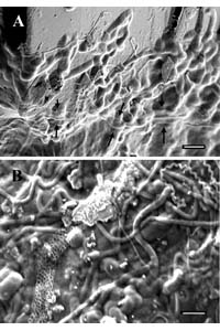 (A) Edge of a microbial mat attached to a mineral surface. The diameter of the smallest filaments is about 70 nm, the bigger ones of approximately 180 nm. Integrated in the mat is a ribbon starting at the left lower edge and continung horizontally (arrows). The web-structure is hardly recognizable because the meshes are filled with slipped-in material. Except for the middle part, the ribbon again seems to follow an underlying feature. (B) Mat community with clearly distinguishable microbes (rods 1.5 x 0.8 micrometer, cocci 1.1 x 0.7 micrometer, big filaments, spirillae and small filaments (diameter 300, 120 and 60 nm, resp.) and a ribbon (850 nm diameter) with a well-defined web-structure. In this case a "leading feature" underneath is not apparent. Variations of the shape of the ribbons 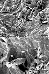 Variations of the shape of the ribbons: (A) The ribbon beginning with its normal appearance (lower right) is further up folding over a sharp ridge (arrow 1). After a 90 degree bend it curls on the upslope of a triangular pebble, and even more at the downslope, deflating to a compact strand with a diameter of 150 nm (arrow 2). Further to the left it resumes its normal structure. In the center (arrow 3), a part of the ribbon has totally collapsed; the remnants are partly discernible as a string of beads (arrow 4). (B) Twisting of the web (arrow 1, 2) leads to the formation of clusters (arrow 3), with bead-like components recognizable. The deflating area at left (4) arises from a folded (4a) and a “filled-up”ribbon (arrowhead). Bars = 1 micrometer Jeany |
|
|
|
Post by jeany on Jul 26, 2009 11:19:58 GMT -5
 Twisting, folding and piling up as huge clusters The ribbon on a smooth mineral surface (A, arrow) begins to fold up after a downward turn on a rougher surface, and curls up to two connected clusters, with remnants of the web-structure visible between these piles (arrowhead). Within the crumpling clusters (B = detail from A) hexagons of various size are recognizable (arrows), but even more pronounced in (C). Short chains and longer loop-forming strings are characteristic for the cluster in (D). 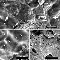 Bead-like substructures and the formation of loops Bead-like substructures and the formation of loops: (A) The bulk (center, right) consists of small strings (+/- 30 nm diameter) composed of tiny beads (better recognizable as individual rows or chains, arrows 1 = ~150 beads = 4.5 micrometer, 2 = ~84 beads = 2.5 micrometer). The strings show a tendency to form loops, frequently with an interspace close to that of the web-structures (arrows 3, 4, as examples); (B) beads at the upper rim, as part of the segments, within the interspace of a web, and very short strings; right foreground: clusters of nanobacteria (230 x 70 nm); (C) segments of an inflated ("puffed-up") web consisting of individual and lined-up beads (arrows); the beads also appear in the interspace of the ribbon structure. Jeany |
|
|
|
Post by jeany on Jul 26, 2009 11:20:54 GMT -5
New life form may be a great find of the century
Nanobes
THE JURY is still out until the DNA evidence is presented, but the case fornanobes being living things is getting stronger by the month.
Up till now, critics have argued they couldn't have been bacteria, on thegrounds that there were no living examples of nano-sized cells on earth -but now, almost certainly, there are. Other theories - that nano-bacteria may be responsible for kidney stones and be involved in processes such as rusting and the greening of copper - are now also being taken more seriously, and the door is opening onto a whole new area of knowledge.
A further impetus came when she noticed that containers and equipment in thelaboratory were being "colonised", and realised that whatever she had foundwas growing, becoming visible to the naked eye within two to three weeks.
The tests showed the nanobes fulfilled the following criteria to qualify as life: -
* their colonies grew spontaneously;
* they contained genetic material (DNA);
* they were composed of biological material such as carbon, oxygen and nitrogen;
* ultra-thin sections showed an outer layer or membrane that could represent a cell wall, surrounding a possible cytoplasm and nuclear area.
Further, they tested a variety of plausible non-biological explanations forthe nanobes, gradually discounting materials such as crystalline materials, carbonates, fullerenes, carbon nano-tubes and non-living polymers.
Dr Uwins and her team have now submitted another paper for publication,which provides further evidence for nanoscopic life.
She says nanobes show a striking morphological similarity to fungi, only on amuch reduced scale.
"They are very tough structures," said Dr Uwins. "Usually, if you're going to process a biological specimen for a scanningelectron microscope, you put it through fixation and dehydration to maintainits structure, but the nanobes will go in and out of microscopes andwithstand the vacuum and the electron beam and the X-rays.
"This is where the debate comes in over the Martian mineralised bacteria,because it suggests they can withstand space travel."
One critic of the research has pointed out that nanobes could be thedegraded remains of organic matter from oil.
Jeany
|
|
|
|
Post by jeany on Jul 26, 2009 11:24:54 GMT -5
www.astrobio.net/index.php?option=com_exclusive&task=detail&id=983 Some claim they are a new life form responsible for a wide range of diseases. Now a team of doctors has entered the fray surrounding the existence or otherwise of nanobacteria. After four years' work, the team, based at the Mayo Clinic in Rochester, Minnesota, has come up with some of the best evidence yet that they do exist. But in 1998 the debate took a different twist when Olavi Kajander and Neva Ciftcioglu of the University of Kuopio in Finland claimed to have found nanobacteria, surrounded by a calcium-rich mineral called apatite, in human kidney stones. Kajander and Ciftcioglu, however, insisted that they had observed the nanoparticles self-replicating in a culture medium and claimed to have identified a unique DNA sequence. After studying nanoparticles found in saliva, his team published a paper in 2000 claiming that the DNA detected by the Finnish team was a contaminant from a normal bacterium. "They talk about 'self-propagating apatite' This suggests that RNA is being produced in the particles, the team says. However, even apatite crystals alone seemed to absorb some uridine, though not as much as the self-replicating particles. And when the Mayo team doused their tissue samples with an antibody that Nanobac claims binds to a protein unique to nanobacteria, they found it bound to diseased tissue, even when the calcium was washed away, but not to healthy tissue. Jeany
|
|
|
|
Post by jeany on Jul 26, 2009 11:26:18 GMT -5
ammin.geoscienceworld.org/cgi/content/abstract/83/11-12_Part_2/1541Nanobes have cellular structures that are strikingly similar in morphology to Actinomycetes and fungi (spores, filaments, and fruiting bodies). Ultra thin sections of nanobes show the existence of an outer layer or membrane that may represent a cell wall. Nanobes show a positive reaction to three DNA stains, [4,6-diamidino-2 phenylindole (DAPI), Acridine Orange, and Feulgen], which strongly suggests that nanobes contain DNA. Nanobes are communicable and grow in aerobic conditions at atmospheric pressure and ambient temperatures. Jeany
|
|
|
|
Post by jeany on Jul 26, 2009 11:27:53 GMT -5
www.panspermia.org/whatsne30.htmJust balls of protein? This is Nature's suggested description for nanofossils like the ones seen in a meteorite from Mars seven years ago. At that time NASA reported that evidence in a meteorite (designated ALH 84001) pointed to former life on Mars. NASA's team, headed by David S. McKay, cited four lines of evidence, one of which was the presence of fossils resembling nanobacteria — later renamed "nanobes." They are too small to contain the genome of a cell, but nanobes are apparently biological products, because they have DNA, protein, and a means to be multiplied, we now know. Nanobes may be the distress byproducts of whole bacteria, for example. "Schieber and Arnott dipped pieces of bean, squid and beef into the muck scooped from a pond bed, coating them in a range of natural bacteria, and then buried the samples under clay in a water tank. Over the next fortnight the researchers regularly studied samples under the microscope. The tissues, they found, became covered in spherical blobs of organic matter." 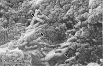 Jeany
|
|
|
|
Post by jeany on Jul 26, 2009 11:36:03 GMT -5
Nanobacteria and Nanobes- Are They Alive? serc.carleton.edu/microbelife/topics/nanobes/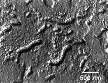 High resolution TEM image of potential nanofossils found in Mars meteorite ALH84001. Nanobacteria are thought to have been found in human blood and may be related to health issues such as the formation of kidney stones due to their biomineralizaton processes. This has been met with some resistance, as some argue that this biomineralization is caused by the nucleation of non-living biological molecules. www.pnas.org/content/97/21/11511.full?sid=bafb7b53-722f-433a-a78a-b6aa87c52786In 1998, Finnish scientists Olavi Kajander and Neva Ciftcioglu published a paper in the Proceedings of the U.S. National Academy of Science about the isolation, culturing, and partial characterization of ribosomal RNA (rRNA) for nanobacteria in human and cow blood and commercial blood serum. Their cultured nanobacteria were made up of apatite, a mineral composed of calcium and phosphate, which is found in teeth and bones. * Organisms may be smaller than previously thought. Proceedings from a workshop (more info) hosted by the National Academy of Sciences in 1998 suggested that the simplest organism would need to be 0.2-0.3µm (200-300 nm) in diameter to hold the molecular pieces necessary for life (e.g. RNAs, ribosome, protein). * If nanofossils exist in Martian meteorites, this may indicate the existence of water at one point in Mars' history. According to current knowledge, water is thought to be essential for life to exist. * Nanobacteria may mediate processes currently thought to be controlled by inorganic chemical reactions, such as low-temperature precipitation of dolomite, oxidation of iron, and the formation of clay minerals (Folk, 1993). (more info) * Nanobacteria may build essential parts of larger organisms or play a role in disease in the same organisms. Biomineralization could result in the formation of bones, shells, teeth, and kidney stones, and arterial plaque. Jeany |
|
|
|
Post by jeany on Jul 26, 2009 11:38:14 GMT -5
Nanobes - Could these primitive life forms be an answer to our own very existance? analogik.com/article_sci_nanobes.aspThey have hollow, membrane bound structures that are most likely composed of C, O and N. Whilst morphologically distinct, nanobes are in the same size range as the controversial nanobacteria described by others in a variety of different rock types and in the Martian meteorite ALH84001. Current and future research will focus on the establishment of axenic cultures for analysis of growth rates and for determining the nature of their genetic material. 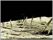 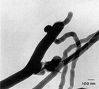 Ten times smaller than any living creature... with a bizarre appetite for plastic and with relatives that may have come from Mars... Nanobes are very strange creatures indeed. What? Nanobes..appetite for PLASTIC?? Jeany |
|
|
|
Post by jeany on Jul 26, 2009 11:39:39 GMT -5
www.chrisamillion.com/news/2004/11/nanobacteria.htmlAnother interesting observation about the nanobes it that they appear to eat plastic! The petri-dish in which the original samples were taken from was completly grown over with nanobes and the plastic was etched, this means that the nanobes not only challenge everything we know about life but they could have a major financial benefit, billions of dollars, how many wasted plastic cups are there in the world! Could it be that Nanobacteria in our water and soil is eating plastic from environmental contamination and it's able to 'combine' together? Is this the connection between Nano..Bacteria..Environmental toxins...and the so called 'plastic fibers'? hmm... Jeany
|
|
|
|
Post by jeany on Jul 26, 2009 11:42:19 GMT -5
Watch and listen!
|
|
|
|
Post by jeany on Jul 26, 2009 11:43:38 GMT -5
Another aspect to consider: www.medicalnewstoday.com/articles/22413.phpThe radical theories about nanobacteria - micro-organisms considerably smaller than ordinary bacteria - in clouds are published in two recent articles in the Journal of Proteome Research by Dr Andrei P. Sommer of the University of Ulm, Germany, and Professor Chandra Wickramasinghe of Cardiff University, UK. They say nanobacteria are now accepted as being widely prevalent in the terrestrial environment and that their evidence is compelling for the existence of these nano-organisms, even in the stratosphere. In humans, nanobacteria have now been identified on four continents, they add. Experiments have shown that nanobacteria are excreted from the body in urine and their dispersal from the ground into the atmosphere and stratosphere appears to be inevitable. This happens because nanobacteria, lifted from the ground by winds, could transit between the high humidity region of the clouds and the relatively dry inter-cloud regions, leading to oscillations between a dormant state and one of activation," explained Professor Wickramasinghe. "Remnants of a sticky protein (slime) coating nanobacteria makes them act as extremely efficient cloud condensation nuclei, with a tendency to aggregate to clusters upon contact." The contribution of nanobacteria to pathogenic bioaerosols, in the view of the authors, must overwhelm all other types of biological particles in the atmosphere. Jeany |
|


