|
|
Post by jeany on Nov 13, 2009 20:44:13 GMT -5
Zygomycosis is the broadest term to refer to an infection caused by fungi of the zygomycetes order.
Rhizopus belongs to the Zygomycetes Family.
Zygomycosis can refer to mucormycosis (after Mucorales), phycomycosis (after Phycomycetes) and basidiobolomycosis (after Basidiobolus), rare yet serious and potentially life-threatening fungal infections, usually affecting the face or oropharyngeal cavity.
Zygomycosis is often caused by common fungi which can be found in soil and decaying vegetation.
While most individuals are exposed to the fungi on a regular basis those with immune disorders are more prone to an infection.
As such, it usually infects those who are immunocompromised.
Occasionally, when caused by Pythium or similar fungi, the condition may affect the gastrointestinal tract or the skin.
It usually begins in the nose and paranasal sinuses and is one of the most rapidly spreading fungal infections in humans.
Pathogenic Zygomycosis is caused by species in two orders: Mucorales and Entomophthorales,
Order Mucorales (mucormycosis)
Family Mucoraceae
Absidia (Absidia corymbifera)
Apophysomyces (Apophysomyces elegans)
Mucor (Mucor indicus)
Rhizomucor (Rhizomucor pusillus)
Rhizopus (Rhizopus oryzae)
Family Cunninghamellaceae
Cunninghamella (Cunninghamella bertholletiae)
Family Thamnidiaceae
Cokeromyces (Cokeromyces recurvatus)
Family Saksenaeaceae
Saksenaea (Saksenaea vasiformis)
Family Syncephalastraceae
Syncephalastrum (Syncephalastrum racemosum)
Order Entomophthorales (entomophthoramycosis)
Family Basidiobolaceae
Basidiobolus (Basidiobolus ranarum)
Family Ancylistaceae
Conidiobolus (Conidiobolus coronatus/Conidiobolus incongruus)
Zygomycosis frequently involves the sinuses, brain, or lungs as the sites of infection.
While oral or cerebral zygomycosis are the most common types of the disease, this infection can also manifest in the gastrointestinal tract, skin, and in other organ systems.
Affected skin may appear relatively normal during the earliest stages of infection.
This skin quickly progresses to an erythemic (reddening, occasionally with edema) stage, before eventually turning black due to necrosis
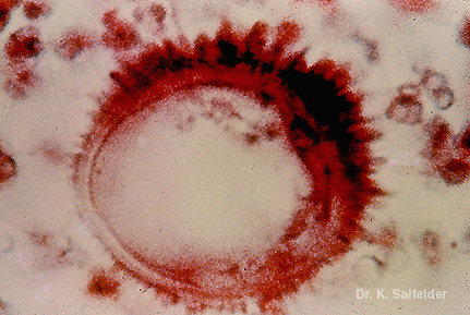
Diagnosis is often difficult because basidiobolomycosis is a rare disease and therefore often not recognised.
The lesions often look like tumours rather than infection, so often no sample is sent for microbiology, however, the histopathology is characteristic: the "Splendore-Hoeppli phenomenon" describes the presence of fungal hyphae (which may exist only as faded streaks on the film) surrounded by eosinophilic material.
Eosinophilia:
Hypereosinophilic syndrome
Parasitic infections (intestinal helminthiasis)
Allergic disorders (including eosinophilic esophagitis)
Some drug reactions, e.g. DRESS syndrome
Cholesterol embolization
Churg-Strauss syndrome
Some forms of chronic myeloid leukaemia
Hodgkin's lymphoma
Gleich's syndrome
Addison's disease
Clonorchis sinensis, a type of flatworm
Eosinophilia-myalgia syndrome caused by contaminated tryptophan supplements
In patients with deep involvement, the eosinophil count may be raised, falsely suggesting a parasitic infection.
Zygomycosis also has similar symptoms to other diseases including anthrax, aspergillosis and cellulitis.
Jeany
|
|
|
|
Post by jeany on Nov 13, 2009 20:46:37 GMT -5
Mucocutaneous Splendore-Hoeppli phenomenonThis is very interesting: www.ncbi.nlm.nih.gov/pubmed/18976399Splendore-Hoeppli phenomenon (asteroid bodies) is the in vivo formation of intensely eosinophilic material (radiate, star-like, asteroid or club-shaped configurations) around microorganisms (fungi, bacteria and parasites) or biologically inert substances. ...It also discusses the mucocutaneous infections and the non-infective diseases associated with it. Available studies indicate that several mucocutaneous infections can generate Splendore-Hoeppli reaction... ...The fungal infections include sporotrichosis, pityrosporum folliculitis, zygomycosis, candidiasis, aspergillosis and blastomycosis. The bacterial infections include botryomycosis, nocardiosis and actinomycosis. The parasitic conditions include orbital pythiosis, strongyloidiasis, schistosomiasis and cutaneous larva migrans. ...it is thought to be a localized immunological response to an antigen-antibody precipitate related to fungi, parasites, bacteria or inert materials. The characteristic formation of the peribacterial or perifungal Splendore-Hoeppli reaction probably prevents phagocytosis and intracellular killing of the insulting agent leading to chronicity of infection. Jeany
|
|
|
|
Post by kammy on Nov 14, 2009 12:07:11 GMT -5
Splendore-Hoeppli Looking at the microscopic images to see what it looks like: Yes, Jeany, we need to look closer at this aspect. The more KNOWN aspects that we can report to our doctors as places to look, the better. |
|
|
|
Post by jeany on Nov 14, 2009 12:22:38 GMT -5
Dipteran-active uracil derivative - Uracil Pesticide
Here's the whole mix:
As defined herein, "a Bacillus related pesticide" is a Bacillus (e.g., Bacillus thuringiensis or Bacillus subtilis) strain, spore, or substance, e.g., protein or fragment thereof, with activity against or which kill pests or provides plant protection against a pest; or a microorganism capable of expressing a Bacillus gene encoding a Bacillus protein or fragment thereof with activity against or which kills pests or provides plant protection against a pest (e.g., Bacillus thuringiensis delta-endotoxin), and an acceptable carrier.
The pest may be, for example, an insect, a nematode, a mite, or a snail.
A microorganism, capable of expressing a Bacillus gene encoding a Bacillus protein or fragment thereof with activity against or which kill pests or provides plant protection against a pest, inhabits the phylloplane (the surface of the plant leaves), and/or the rhizosphere (the soil surrounding plant roots), and/or aquatic environments, and is capable of successfully competing in the particular environment (crop and other insect habitats) with the wild-type microorganisms and provide for the stable maintenance and expression of a Bacillus gene encoding a Bacillus protein or fragment thereof with activity against or which kill pests.
Examples of such microorganisms include, but are not limited to, bacteria, e.g., genera Bacillus, Pseudomonas, Erwinia, Serratia, Klebsiella, Xanthomonas, Streptomyces, Rhizobium, Rhodopseudomonas, Methylophilius, Agrobacterium, Acetobacter, Lactobacillus, Arthrobacter, Azotobacter, Leuconostoc, Alcaligenes, and Clostridium; algae, e.g., families Cyanophyceae, Prochlorophyceae, Rhodophyceae, Dinophyceae, Chrysophyceae, Prymnesiophyceae, Xanthophyceae, Raphidophyceae, Bacillariophyceae, Eustigmatophyceae, Cryptophyceae, Euglenophyceae, Prasinophyceae, and Chlorophyceae; and fungi, particularly yeast, e.g., genera Saccharomyces, Cryptococcus, Kluyveromyces, Sporobolomyces, Rhodotorula, and Aureobasidium.
The Bacillus related protein may be selected from the group including, but not limited to, CryI, CryII, CryIII, CryIV, CryV, and CryVI.
The chemical pesticide may be, for example, an insect growth regulator such as diflubenzuron, a carbamate such as thiodicarb and methomyl, an organophosphate such as chlorpyrifos, a pyrethroid such as cypermethrin and esfenvalerate, inorganic fluorine such as cryolite, and a pyrrole.
The entomopathogenic virus may be a baculovirus, e.g., Autographa californica nuclear polyhedrosis virus (NPV), Syngrapha falcifera NPV, Cydia pomonella GV (granulosis virus), Heliothis zea NPV, Lymantria dispar NPV, Orgyia pseudotsugata NPV, Spodoptera exigua NPV, Neodiprion lecontei NPV, Neodiprion sertifer NPV, Harrisina brillians NPV, and Endopiza viteana Clemens NPV.
Jeany
|
|
|
|
Post by jeany on Nov 14, 2009 12:52:16 GMT -5
I found this very interesting...
Contaminated sewage sludge used as a fertilizer on food crops? FUNGUS IN SLUDGE - BIOSOLIDS - natural borne killersdeadlydeceit.com/fungus_sludge.htmlAn example of the deliberate production of ignorance is the 1994 EPA and Water Environment Federation (WEF) public relations program to change public perception about the danger to human health of sludge promoted as a fertilizer and soil amendment. Under the banner of sound science, the industry produced research about everything except disease organisms -- and -- fungus in sludge - biosolids was completely ignored.
The real question is: how can the laboratories in Canada and the United States understand the danger from exposure to fungus and yet, the government agencies from both nations remain ignorant of the facts. Current FDA Requirements for Spore-Forming Microorganisms which are ignored in sludge - biosolids handling.The ignorance was maintained in 1981 when USDA, FDA, and CDC joined EPA in creating a national policy to use sewage sludge as a fertilizer on food crops. Even though these agencies claim they didn’t understand the health
significance of fungus in sludge, the policy of ignorance was expanded in 1989 to distributed and marketed
sludge as a fertilizer and soil amendment. The expanded policy of ignorance was based on a 1988 compost study by the Los Angeles County Sanitation District who had to dispose of huge quanities of sludge.n the 1988 EPA study "Occurrence of Pathogens in Distribution and Marketing Municipal Sludges," William A. Yanko, County Sanitation Districts of Los Angeles County, California states: “A number of pathogenic or allergic fungi can be isolated from sludge. These include yeasts, such as certain specious of Candida, Cryptocuccus and Trichosporon, and pathogenic members of some filamentous genera, such as Aspergillus, Phialophora, Geotrichum, Trichophyton and Epidermophyton.”" Aspergillus fumigatus, an opportunistic pathogen to individuals with pulmonary problems and a strong allergen to many, may proliferate in some composting systems. This may be a consideration when selecting prospective composting sites. The general consensus, however, is that fungi in treated sludges present a minimal hazard. With the exception of the aspergilli, little work has been done to define the relationships of fungi in polluted environments or sludges. The significance, if any, of fungal types and diversity in compost is unknown.” "Dermatophytic fungi [cause skin infections] , for example, may be present in sludge at detectable levels. It is not known if the common dermatophytes can survive or proliferate in sludges. Conventional thought considers the dermatophytes to be parasitic, although there is evidence that some dermatophytes live a saprophytic existence. Adding large numbers of these organisms to home soils would be undesirable."For the past 20 years EPA, USDA and WEF studies have focused on chemicals and non-disease causing bacteria in an effort to prove sludge used as a fertilizer and soil amendment is safe.
EPA has admitted: 1) that its risk
assessment did not consider any chemical in sludge to cause cancer (Los Angeles employees couldn't identify
most chemicals in EPA study); 2) that it did not do a risk assessment for disease causing organisms; and 3) it
pleads at least semi-ignorance about the danger to health of fungus in sludge - biosolids.EPA’s Alan Rubin (retired) has claimed to be the genius behind the EPA’s 503 sludge use and disposal policy
guideline. Rubin has been the public face of EPA on sludge and its chief salesman during the public relations campaign of lying to the public.. Rubin’s biggest argument for the safety of sludge use was that it was safe as long as the laws and regulations were followed. However, his biggest omission to the public and politicians was that 503 was based on exclusions in the environmental laws. While EPA claims there are no victims, but Cornell has a limited list. Under a freedom of information request, Rubin wanted to charge Helene Shields $40,000.00 dollars in copy and labor cost for copies of sludge victims complaints. It would appear that Rubin also forgot a key definition in his guideline that states exposure to any of the pollutants in sludge (i.e. organic, inorganic chemical, disease organisms or a combination of them), through air, water or food could cause death, disease, cancer, etc.. Nevertheless, since his retirement, Rubin has become a consultant for the sludge industry promoting the safety of sludge use. However, today the public has a better understanding of science. Rhizopus is fungus found in sludge - biosolids compost, soil, decaying fruit and vegetables, animal feces, and old bread. Rhizopus spp. may cause serious (and often fatal) infections in humans. Some species are plant pathogensRhizopus cause infections in the rhino (eye, nose, throat) -facial (includes dental) - cranial (brain and spine) area, lungs, gastrointestinal tract, skin, vessel (arterial) invasion resulting in embolization and necrosis (death) of surrounding tissue, Rhinocerebral disease in acidotic patients usually results in death, often within a few days, stomach, ileum, and colon, heart, bone, kidneys, bladder, trachea, and mediastinum, ((the part of the thoracic cavity between the lungs that contains the heart and aorta and esophagus and trachea and thymus), mouth palate, necrotic (dead tissue) ulcers, cover for nerve fibers, may include prostatitis, benign prostatic hyperplasia (abnormal development (of organs or cells)), urogenital cancers, premature ejaculation and erectile dysfunction, The potential problems to be expected from sludge compost according to EPA:: Survival and presence of primary pathogens in the product.Composting is not a sterilization process and a properly composted product maintains an active population of beneficial microorganisms that compete against the pathogenic members. Under some conditions ,explosive regrowth of pathogenic microorganisms is possible.Dispersion of secondary pathogens such as Aspergillus fumigatus, particulate matter, other airborne allergens. While healthy individuals may not be affected, immunocompromised individuals may be at risk.The spores of A. fumigatus counts at composting facilities are high, and-- persons handling composted biosolids being exposed to these spores is also high (Epstein, 1998).These organisms can potentially invade a normal, healthy human being and produce illness or debilitation. Jeany |
|
|
|
Post by jeany on Nov 14, 2009 13:17:29 GMT -5
I was thinking about how our hair seems to be infected, degraded and found this site. Characterization of a new keratin-degrading bacterium isolated from deer furtinyurl.com/yfon3phA keratin-degrading bacterium was isolated from soil containing deer fur. ...Stenotrophomonas nitritireducens. Hence, this new bacterium was designated as Stenotrophomonas sp. D-1. Jeany |
|
|
|
Post by jeany on Nov 14, 2009 13:39:35 GMT -5
|
|
|
|
Post by jeany on Nov 14, 2009 13:43:53 GMT -5
Characterization of Flagella Produced by Clinical Strains of Stenotrophomonas maltophilia www.cdc.gov/ncidod/EID/vol8no9/01-0535.htmStenotrophomonas (formerly Pseudomonas and Xanthomonas) maltophilia is a widespread environmental microorganism that has become an important opportunistic pathogen associated with nosocomial colonization and infection. These organisms have been recovered from water faucets, water traps, respirometers, sinks, suction catheters, and occasionally, from cultures of the hands of hospital personnel. Infection and colonization of implantable medical devices such as catheters and intravenous cannulae represent a major risk for hospitalized patients. S. maltophilia can cause septicemia, endocarditis, conjunctivitis, mastoiditis, meningitis, postoperative wounds, abscesses, urinary tract infections, and pneumonia.  Ultrastructural analysis of Stenotrophomonas maltophilia adhering to plastic. (A) Scanning electron micrographs showing the tight adhesion of SMDP92 to the plastic surface. (B) Structures resembling flagella seem to be protruding and interconnecting bacteria (arrowheads) or connecting bacteria to the plastic (arrows). (C) In addition to the flagellalike filaments (arrowheads), high-power magnification shows the presence of thin fibrillar structures connecting bacteria to the abiotic surface. Bars: A 10 mm, B 1 mm, C 2 mm. Jeany |
|
|
|
Post by jeany on Nov 14, 2009 16:03:47 GMT -5
www.isradiology.org/tropical_deseases/tmcr/chapter6/clinical15.htmZygomycosis is the general term for an infection caused by pathogenic zygomycetes of the orders Murocales and Entomophthorales. Mucormycosis is a systemic fungal infection caused the genera Rhizopus, Mucor, or Absidia. Less often it is due to Cunninghamella or Saksenaea. Entomophthoromycosis is infection caused by one of the Entomophthorales, usually Basidiobolus haptosporus or Conidiobolus coronatus, and very rarely Conidio bolus incongruus. These infections have epidemiological, clinical, and pathological differences from mucormycosis. It seems very possible that clinical and imaging descriptions have not made a clear differentiation, which may be more important therapeutically than for other reasons. Zygomycetes are found worldwide: infections have been reported from the Indian subcontinent, Africa, South America, Asia, Europe, and the United States. The zygomycetes are the common molds found on bread, cheese, and fruit. They are also present in decaying vegetation. Mucormycosis seldom occurs in healthy individuals, although some have been infected while apparently otherwise healthy. It is most common in patients who are immunocompromised for any reason, including diabetes, renal failure, leukemia, the reticulosis, and malnutrition. Mucormycosis is rare in AIDS patients, but both may occur in intravenous drug users. Skin and soft tissue infection has followed local trauma. Intravenous drug abuse is another common factor. Several infections have been linked with contaminated adhesive bandages/tape which have been left in place too long so that the underlying skin has become damaged. Organ transplantation and clinical immunosuppression for any reason increase the risk of infection. The Entomophthorales are found in the alimentary tract of many amphibians, reptiles, horses, dogs, and chimpanzees but few are clinically infected. A significant difference which affects pathogenicity is that, unlike the members of the Mucorales, the organisms of the Entomophthorales tend not to invade the wall and lumen of blood vessels (although a few cases of disseminated infection have been recorded). Because vascular invasion is not a feature of the entomophthomycoses, neither septic thrombosis nor infarction and the subsequent necrosis are observed as with mucormycoses. The entomophthomycosis tend to be characterized by indolent local infections, in contrast to the usually quickly disseminating mucormycosis. Entomophthora coronata almost always affects only the upper respiratory tract. The hyphae of entomophthoromycoses are septate, while those of mucormycetes are nonseptate; in addition, the hyphae in entomophthomycoses often are enclosed by an eosinophilic sheath (Splendore-Hoeppli phenomenon) not seen in the mucormycoses In other respects the infections are similar, with central abscesses surrounded by numerous eosinophils. Biopsy is the best way to establish the diagnosis because serology is unreliable. Jeany
|
|
|
|
Post by kammy on Nov 16, 2009 18:42:01 GMT -5
Someone told me to look at self-cleaning nanotubes, I find this information interesting: tinyurl.com/yjpq8vuFebruary 12th, 2008 Nanotechnology-based self-cleaning fabrics In one of their experiments, they poured red wine upon pristine and nanotechnology-coated wool. After 20 hours, their coated fabric ’showed almost no signs of the red stain, whereas the untreated fabric remained deeply stained.’ “The coating, which is non-toxic, can be permanently bonded to the fibre and does not alter its texture and feel, they note, so a silk tie would still feel silky. The tricky part of the research was finding a way to bind the keratin to the titanium dioxide, says Daoud. ‘Applying a ceramic inorganic material to organic fibres, in particular keratin protein fibres such as wool, silk, hemp, and spider silk, remained a challenge.’” For more information, this research has been published online by Chemistry of Materials, an American Chemical Society publications under the title “Self-Cleaning Keratins” on January 23, 2008. “Keratins, a class of biologically fibrous proteins, are tough and insoluble due to the formation of adjacent peptide bond that allow close alignment of the sulfur-containing amino acid constituent, cysteine, enabling the formation of disulfide bridges, cross-links, that confer rigidity and thermal stability to keratinous materials, which make them an important class of fibrous materials. Keratins are a type of natural protein and the main structural constituents of animal tissues. Keratinous protein fibers such as wool, silk, hemp, and spider silk find numerous applications such as insulation, tires, and strong fibers in addition to textiles.” “In this contribution, self-cleaning keratin fibers have been realized following a bottom-up nanotechnology approach in which anatase nanocrystals of titanium dioxide are formulated and carefully applied to the fibers via a near room temperature sol–gel process in order to maintain their intrinsic properties while conferring self-cleaning properties and self-protection against UV degradation. This may enable wider utilization of these natural fibers.” Sources: American Chemical Society, February 6, 2008; Roger Highfield, The Telegraph, UK, February 11, 2008; and various websites |
|
|
|
Post by imblownaway on Nov 16, 2009 21:17:01 GMT -5
Someone told me to look at self-cleaning nanotubes, I find this information interesting: tinyurl.com/yjpq8vuFebruary 12th, 2008 Nanotechnology-based self-cleaning fabrics In one of their experiments, they poured red wine upon pristine and nanotechnology-coated wool. After 20 hours, their coated fabric ’showed almost no signs of the red stain, whereas the untreated fabric remained deeply stained.’ “The coating, which is non-toxic, can be permanently bonded to the fibre and does not alter its texture and feel, they note, so a silk tie would still feel silky. The tricky part of the research was finding a way to bind the keratin to the titanium dioxide, says Daoud. ‘Applying a ceramic inorganic material to organic fibres, in particular keratin protein fibres such as wool, silk, hemp, and spider silk, remained a challenge.’” For more information, this research has been published online by Chemistry of Materials, an American Chemical Society publications under the title “Self-Cleaning Keratins” on January 23, 2008. “Keratins, a class of biologically fibrous proteins, are tough and insoluble due to the formation of adjacent peptide bond that allow close alignment of the sulfur-containing amino acid constituent, cysteine, enabling the formation of disulfide bridges, cross-links, that confer rigidity and thermal stability to keratinous materials, which make them an important class of fibrous materials. Keratins are a type of natural protein and the main structural constituents of animal tissues. Keratinous protein fibers such as wool, silk, hemp, and spider silk find numerous applications such as insulation, tires, and strong fibers in addition to textiles.” “In this contribution, self-cleaning keratin fibers have been realized following a bottom-up nanotechnology approach in which anatase nanocrystals of titanium dioxide are formulated and carefully applied to the fibers via a near room temperature sol–gel process in order to maintain their intrinsic properties while conferring self-cleaning properties and self-protection against UV degradation. This may enable wider utilization of these natural fibers.” Sources: American Chemical Society, February 6, 2008; Roger Highfield, The Telegraph, UK, February 11, 2008; and various websites Interesting It reminded me of something. In the very early stages I was so alarmed at my hair moving. Not just moving but moving with intent. I was so scared. I remember spraying it down with hairspray.A ton of it. Going for that plastered down look.  Within a short time my hair felt like I hadn't sprayed it at all. I mean soft clean hair.  I don't know what the hell I have. I just know its bizarre and I want to get well. |
|
|
|
Post by kammy on Nov 17, 2009 8:15:23 GMT -5
Someone told me to look at self-cleaning nanotubes, I find this information interesting: tinyurl.com/yjpq8vuFebruary 12th, 2008 Nanotechnology-based self-cleaning fabrics In one of their experiments, they poured red wine upon pristine and nanotechnology-coated wool. After 20 hours, their coated fabric ’showed almost no signs of the red stain, whereas the untreated fabric remained deeply stained.’ “The coating, which is non-toxic, can be permanently bonded to the fibre and does not alter its texture and feel, they note, so a silk tie would still feel silky. The tricky part of the research was finding a way to bind the keratin to the titanium dioxide, says Daoud. ‘Applying a ceramic inorganic material to organic fibres, in particular keratin protein fibres such as wool, silk, hemp, and spider silk, remained a challenge.’” For more information, this research has been published online by Chemistry of Materials, an American Chemical Society publications under the title “Self-Cleaning Keratins” on January 23, 2008. “Keratins, a class of biologically fibrous proteins, are tough and insoluble due to the formation of adjacent peptide bond that allow close alignment of the sulfur-containing amino acid constituent, cysteine, enabling the formation of disulfide bridges, cross-links, that confer rigidity and thermal stability to keratinous materials, which make them an important class of fibrous materials. Keratins are a type of natural protein and the main structural constituents of animal tissues. Keratinous protein fibers such as wool, silk, hemp, and spider silk find numerous applications such as insulation, tires, and strong fibers in addition to textiles.” “In this contribution, self-cleaning keratin fibers have been realized following a bottom-up nanotechnology approach in which anatase nanocrystals of titanium dioxide are formulated and carefully applied to the fibers via a near room temperature sol–gel process in order to maintain their intrinsic properties while conferring self-cleaning properties and self-protection against UV degradation. This may enable wider utilization of these natural fibers.” Sources: American Chemical Society, February 6, 2008; Roger Highfield, The Telegraph, UK, February 11, 2008; and various websites Interesting It reminded me of something. In the very early stages I was so alarmed at my hair moving. Not just moving but moving with intent. I was so scared. I remember spraying it down with hairspray. A ton of it. Going for that plastered down look. Within a short time my hair felt like I hadn't sprayed it at all. I mean soft clean hair. I don't know what the hell I have. I just know its bizarre and I want to get well. Yes, it is interesting IBA, there's not much written about this material out there, though and this is a year and a half old. This person thinks this has something to do with our disease... We've seen that there's something happening inside some of our hair shafts - I've only looked at the 'wild' ones... not the others - and see that there's some sort of movement happening inside them... maybe, someone else will look at one of their 'wild' hairs under the microscope and report what they see? |
|
|
|
Post by ruth on Nov 17, 2009 12:46:22 GMT -5
i have had this for 13 years.
i do not have any normal hair left.
it is easy to see the infection within the shaft.
i sent a mass of hair (among lots of other samples)
to mark darrah. it was in the last batch i sent him
and he had just gotten evicted from the lab, so he
reportedly threw them away.
|
|
|
|
Post by kammy on Nov 17, 2009 15:42:35 GMT -5
There's something 'fishy' going on... if any of you get a wild hair to look...
I'm wondering if the movement I saw inside the white hair shaft could be a form of this 'self-cleaning'...?
|
|
|
|
Post by kammy on Nov 18, 2009 9:07:30 GMT -5
I was reading on another site where this person was concerned and asking if Morgellons was contagious. I didn't think it appropriate to copy and bring it here... but, if you view the other sites, you might see it? She mentions in her post that this fellow has Morgellons and that he had captured some of 'them'... and she saw how these entities were 'shape shifters' that changed before her eyes and how they stacked themselves in a 'chain'. I don't know if you noticed that this is what Jeany and I described when we saw the fungus gnat larvae change before our eyes and I put out a photo of two of them stacked on top of each other, they form a quorum sensing chain. I saw this article below in the MSN news today... and have thought - what besides a temperature and moisture change caused these larvae to react in this changing and stacking behavior? It was as if they are afraid of humans, when we standing over them, viewing them... they knew it! We've heard testimonies of people posting about these entities under their skin having a like mind, etc., this article makes me wonder? "Tiny insect brains can solve big problems Some bugs can recognize human faces, count and categorize, studies say www.msnbc.msn.com/id/33974286/ns/technology_and_science-science/By Emily Sohn updated 5:52 p.m. ET, Mon., Nov . 16, 2009 Insects may have tiny brains, but they can perform some seriously impressive feats of mental gymnastics. According to a growing number of studies, some insects can count, categorize objects, even recognize human faces — all with brains the size of pinheads. Despite many attempts to link the volume of an animal's brain with the depth of its intelligence, scientists now propose that it's the complexity of connections between brain cells that matters most. Studying those connections — a more manageable task in a little brain than in a big one — could help researchers understand how bigger brains, including those of humans, work." |
|
|
|
Post by kammy on Nov 18, 2009 16:12:12 GMT -5
|
|
|
|
Post by kammy on Nov 20, 2009 11:57:49 GMT -5
'Miez Miez' (pronounced Meats Meats) is getting to be a big boy... this is gross but, I think it's important to share. We're often worried about giving Morgellons to our pets, I've been sloughing particles and try to be aware of where he is, it's impossible to protect them when there's a large amount of debris coming out of us... I just pray that his immune system is strong and can fight this. I was negligent recently in regard to emptying his litter box... I hate messing with them to begin with, he goes outside and doesn't use it that much - so, it was about a week when I decided to change it yesterday. I was surprised to see that two pieces of his stool had the white, fuzzy, cotton candy fungal growths around them. I asked Jeany to come look as I was emptying it outside... now, I worry that he may have "M", even though he doesn't have any signs of it. I still have a wandering gnat in the house once in a while... she thinks that maybe the gnats took up in his litter box. I'm hoping this is it. I think it's a crying shame that our pets are getting this and our Vets are just as lost as our doctors. Anyway, what I was thinking is that this may be a way to detect if your cat has "M" or not? If you allow their stools to sit for a few days, up to a week - the white fungus should show up...? and it might help the Vets if you took their stool in and let the Vets see this fungus? (Of course, it would be best if you didn't have an environmental gnat issue.) Just saying... We haven't cultured a stool smear yet... I just bet it shows it, also. With the other, easier ways that are more sanitary - this shouldn't be necessary to show fungal growth but might be an interesting way to detect other things? As I was looking at his stool... it reminded me of the fungus that strawberries get. I'm thinking that we shouldn't eat fresh strawberries until we know more... maybe somebody needs to culture a small piece of strawberry? We are the plant, we have a plant fungal disease?... what fungal diseases do strawberries and red grapes get? *I believe our fungus starts out as white, to gray then turns to black/green? |
|
|
|
Post by jeany on Nov 20, 2009 12:41:18 GMT -5
Don't want to talk about it much..it still hurts, but I think Mikey had Morgellons too..my poor baby! Gosh..this damn disease!..sry..
I hope and pray that our little MeatsMeats can fight it off...IF he has Morgellons or perhaps the fungal growth was an environmental cause and the gnat got in there..I hope I'm right...
Jeany
|
|
|
|
Post by kammy on Nov 20, 2009 12:56:30 GMT -5
Ok, Jeany is ON IT! I prodded her... and she looked... and BINGO! we think we have a missing ingredient! and we will continue this over in Frito's thread... "kammy's request" - our worlds have collided here.
|
|
|
|
Post by kammy on Nov 21, 2009 1:49:52 GMT -5
|
|


 Within a short time my hair felt like I hadn't sprayed it at all. I mean soft clean hair.


