|
|
Post by jeany on Oct 15, 2009 11:13:34 GMT -5
This is from a post at MDR about this subject and the parasitic wasp for biocontrol we've been working on:
Quote Kammy:
"So, these Saccharomyces cerevisiae organisms which are used in our food processings... affect us how? What are they doing to the actin and filament production?"
From post # 77
The actin nucleation-promoting activity of S. cerevisiae formins has been localized to the FH1-FH2 domains.
"Saccharomyces" derives from Latinized Greek and means "sugar mold" or "sugar fungus", saccharo- being the combining form "sugar-" and myces being "fungus".
Wasn't it mentioned that the fibers contain Polysaccharides? If I recall right?
ok...so Saccharomyces cerevisiae formins promotes the activity of actin nucleation?
.................................................. .................................................. .........
Ok, from what I understand is that fermented products such as cheese, beer or/and food with Fructose syrup for example lead to high level of sugars (glucose) in our bodies which in the other hand interferes with the production of insulin (pancreas) and other hormone producing glands and also the production of actin filaments?
We have also researched that this virus is/was implicated into this bio insecticidal wasp...and that actin (a protein) is involved in viral movements...
Originally Posted by Kammy
To complement our studies in vivo, we study viral pathogenesis and virus-host interactions at the cell and molecular levels as well.
These studies primarily are focused on the role of actin, one of the most abundant cellular proteins, in these processes.
Actin appears to be involved in viral movement, nucleocapsid morphogenesis, and regulation of the switch from production of one viral phenotype to the other.
Accordingly, dramatic sequential changes take place in the distribution and quantity of microfilaments throughout the course of infection which correlate with different phases of virus replication.
In uninfected cells, microfilaments appear to be associated primarily with the plasma membrane.
Upon infection, correlated with virus uptake and independent of early virus gene expression, thickened cables of microfilament bundles appear.
Next, dependent upon early virus gene expression, microfilaments appear as ventral aggregates.
Finally, and dependent on late gene expression, microfilaments appear within the nucleus where they co-localize with newly synthesized capsid protein during nucleocapsid assembly."
.................................................. .................................................. .........................................
This could explain why so many people have reported that a sugar free diet and changing their eating habits have noticed a reduction of symptoms and/or a 'covering-up' of lesions?
Is a sugar-free diet helpful in relieving symptoms..? I think so..
We need to look further:
...how this parasitic wasp...bioinsecticides...bacteria...fungi...actin filaments.....hormones..sugar/glucose...are all linked together..
Kat
|
|
|
|
Post by jeany on Oct 15, 2009 18:24:38 GMT -5
So, I decide to look at Podospora anserina... Podospora anserina tinyurl.com/yl9hgkkLook at this. Could this be the source of the black specks?  Jeany |
|
|
|
Post by jeany on Oct 15, 2009 18:46:32 GMT -5
Here is another article according sup35 and hsp104:
Protein-folding Diseases and Chaperone Biologyweb.wi.mit.edu/lindquist/pub/ChaperoneBiol.htmlHeat-shock proteins (Hsps) are molecular chaperones that are induced when organisms are exposed to high temperatures and other stresses.
These stresses cause proteins to unfold and potentially aggregate, thereby creating a protein-folding crisis in the cell.
Hsp chaperones help the cell cope with this crisis by binding different types of folding intermediates and interacting with them in different ways. One, Hsp104, has a unique ability to promote the disaggregation of aggregated proteins. This biochemical function is in keeping with its biological function: Hsp104 is not required for normal growth but is required for survival under extreme stress. Homologues of Hsp104 have been found in other fungi, bacteria and plants and appear to function in a similar way.Hsp104 is a member of the important AAA family of proteins. AAA-proteins function to remodel other cellular proteins and thus affect a multitude of biological processes. Their power to remodel substrates lies in their capacity to couple substrate binding to conformational change. Hsp104’s ability to remodel protein structures also plays a critical role in protein conformation-based inheritance. In order to understand how Hsp104 functions in this capacity, we are employing a variety of biochemical and genetic approaches.  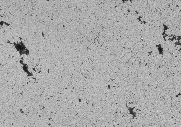 Jeany |
|
|
|
Post by fritolay66 on Oct 16, 2009 8:25:14 GMT -5
Prions.
|
|
|
|
Post by jeany on Oct 16, 2009 9:25:24 GMT -5
Hello all! This is from my blog morgellons2.wordpress.com/ and I would like to know what you think about it..any comments, anyone? Urasil - The source of the fibers?A fellow Morgellons Researcher is working on Micelles and how they are seen in several samples of Morgellons sufferers and how Micelles have the characteristic to form a gel network of fibrils which look highly similar to Morgellons fibers. Interesting was the fact that Micelles are used in water remediation and contain Nanoparticles which interact with chemical substances such as antibiotics. After entering the keywords – Micelles – fibrils – water – I came to a site that is stating and actually showing the main source of the Morgellons fibers in interaction with urasil, water and the ‘creation’ of cellulose fibrils. Researchers and scientists working on Morgellons Disease have revealed that the fibers contain cellulose. I found it very astonishing as I looked at the images which I believe look very similar to Morgellons fibers. Here is a excerpt from this site: Based on the studies of Erwin Schroedinger, we now know that about 5000 nucleotides are lost from the DNA of each human cell by spontaneous hydrolysis due to thermal fluctuations (Schroedinger: 1945 and Alberts: 1983). Furthermore, it is known that some of the deamination products of the DNA bases, such as uracil, hypoxanthine and xanthine are released into the intracellular water. This study focuses on these three DNA deamination products and their behaviour when and if they are not fully metabolised in the organism. My findings lead to interesting conclusions, i.e. the uracil base might have played a crucial role in the evolution process, especially in the conversion of abiogenesis into biogenesis. The uracil base which started biogenesis in the waters in the primitive earth conditions, when separated (as a result of heat fluctuations or upon chemical, physical cancerous effects that spoil the genes) from the genome in today’s living organisms and mixed with cell water, develops itself as it starts the universal Phylogenetic tree from zero and enlarges the entropy of the system founded by the genome. In caryoplasm, cytoplasm, intercellular tissue, artery, blood or in organs and tissues it takes various forms and shows miscellaneous developments. 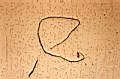 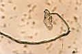 It is neither nucleic acid, gene, virus, bacteria, protozoon, fungus, protein, nor prion, but a preprocaryotic molecule transformed into cellulose capable of giving those appearances. It is neither nucleic acid, gene, virus, bacteria, protozoon, fungus, protein, nor prion, but a preprocaryotic molecule transformed into cellulose capable of giving those appearances. Cellulose making process of uracil base started at the level of molecule, micro fibril, misel and/or fibril, causes the changing of the relationship of the cells with all tissues, organs and systems and the ageing of the system, by escaping the control of the organism’s defence mechanism and by negatively affecting the functions of the cells related to the production of enzymes, hormones, secretes and neuro-secretions etc. Cellulose making process of uracil base started at the level of molecule, micro fibril, misel and/or fibril, causes the changing of the relationship of the cells with all tissues, organs and systems and the ageing of the system, by escaping the control of the organism’s defence mechanism and by negatively affecting the functions of the cells related to the production of enzymes, hormones, secretes and neuro-secretions etc. This next image shows the ‘spikey’ formation that resembles exactly a fellow Morgellons sufferer has shown in microscopical pictures using blood and excreting yellowish liquid samples from his lesions. 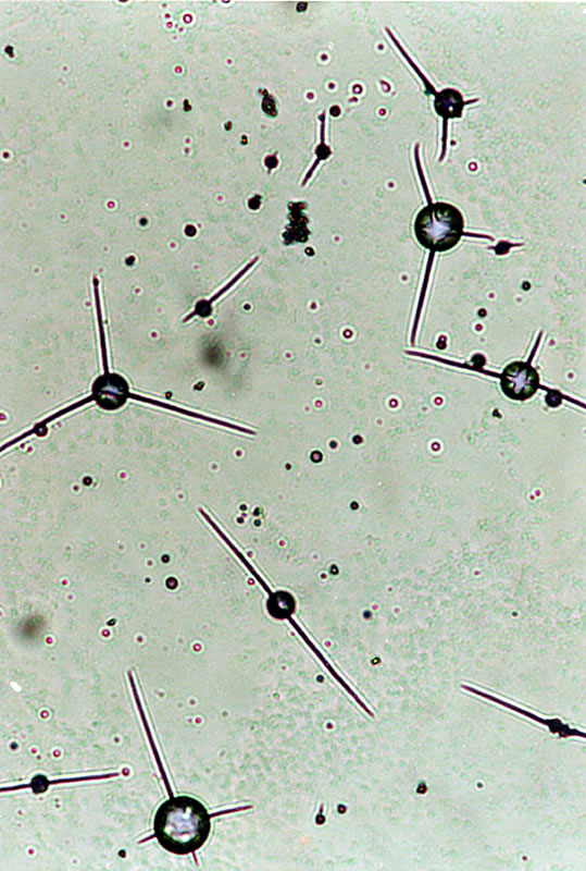 **if you look at the last picture and compare it with the pictures and videos Baraka made I think there is a high similarity to them. www.morgellons-disease-research.com/Morgellons-Message-Board/....post # 1 & 36 Jeany |
|
|
|
Post by camv35s on Oct 16, 2009 10:08:43 GMT -5
Hi jeany regarding the ribbon like fibers I have 100's on slides in plastic siplocks but under the scope alot of mine are motile with undulating movement quite fascinating but the most peculiar quality was its abilty to remain in this state for over a year no oxygen or light water takes a lickin and keeps on tickin,some also have an appendage with a stinger and i equate this with the stings I experiance from time to time used to be more frequent but my regimen of supplements may have slowed that down good site you have best regards camv
|
|
|
|
Post by fritolay66 on Oct 16, 2009 13:42:28 GMT -5
Jeany,
You know, what you guys are talking about kinda reminds me of the baterial enzymatic cleaners, the kinds for toilets and drains and such.
I have a friend in which has a friend, whom was working with those cleaners the other day, and broke out into lesions. I guess this person does not remember if they got it on their skin.
Frito
|
|
|
|
Post by kammy on Oct 16, 2009 14:24:10 GMT -5
Interesting Frito... tell us more? Prions?... you go first... lol... I've got a handful with foods right now, we could use some help out here ya' know? We've got these 'doggies' rounded up... come on let's put some brands on them? Jeany's Uracil find matches researcher Baraka's earlier video of 'Morgellons Forming' 100 percent! WTG, Jeany! Uracil is more than likely in the mix (pesticide related)... Jeany is writing a paper on it, we look forward to... I don't know if you're following my blog postings? Here's one my latest findings about our foods: This is the inside 'meat' of a non-organic banana... just lightly scrape the inside part of a banana on a slide and look at 100x: ![]() my-stuff-dot-com.com/LB/Banana/Meat my-stuff-dot-com.com/LB/Banana/Meat 10 16/10_15_581.jpg[/img] What do you think, Morg related, or not? Speaking of bananas... Jeany and I called Banny yesterday, she is ok... her Internet is down, I'm sure she will be back on soon. Cam, you want to submit some of your pics for comparisons, discussion or analysis?... I noticed Ruth has some good ones... |
|
|
|
Post by camv35s on Oct 16, 2009 16:16:44 GMT -5
hi kammy i would like to submit some pictures but when my computer crashed i lost both hard dives and every other thing I'm on and old computer now waiting for new hard drive installer program I used to post under the name Lucky1 remember me best regards camv.
|
|
|
|
Post by fritolay66 on Oct 16, 2009 18:44:17 GMT -5
If you all know of someone whom has a digital microscope they want to give away, I would be happy to borrow or acquire it. I think I may have lost my job this evening.
Prions.
In the setting in which I was in when I came up with this life changing crap is the hospital setting. I remember someone, perhaps it was Steve Frey in which he saw under his microscope, the organism/whatever transcend the latex glove. Considering the trauma I used to work with, I was basically swimming in patients blood all day.
In this setting prions are a major concern of our surgical instrumentation and of course, the environment itself. Prions, they can withstand radiation, harsh chemicals including bleach, ammonia, you name it. They form Beta sheets on inanimate objects and incorporate proteins into the sheet. Of which then the proteins introduced in the prion setting, change their folding pattern, and their function changes to that of the Beta sheet of the Prion. This process is called an amyloid polymerization.
The reason it is so dangerous in this setting is due to prions introduced into the body, whether through skin contact or surgical contact, is that this protein infection deposits amyloid plaques along the central nervous system. There are many here and myself included in which think the lymphatic system is involved in some form. But the Nervous system also has the same pathways and more in which this infection can invade. The prion infection interferes with tissue structure.
Now lets introduce some modified crap to the mix in which the concern is lateral transfer of DNA not to mention any infections already present in the patient. Infections, latent viruses, unidentified fungal infections, whatever. And by modified crap I am referring to the whole chamiel. Immunizations, GM foods, bacterphages used in our foods, irradiation, water treatments, yadi ya. I don't know but if we look at the last thirty years or perhaps more, the rate of soil organism and insect organism infections in the general population of humans are to say the least, experiencing an increase. Substantial in my opinion, for what its worth.
I guess something else that gets my attention is the fact that there have been many references to a mysterious protein being found amongst us. So just perhaps we have a mystery prion to find. God only knows the combo of it and even experts can't tell you how prions are exactly spread or how they infect.
Another thing to consider is some of these videos in which show the metallics. I remember seeing one video in which the octogonal metallic, unidentified of course, and they peeled it apart in sections. What it looked like to me was some heavy metal in which under the metal was a biofilm..., fungus and bacteria. And then there was the mystery organism. Fungal infections will incorporate metals within the body. They say when treating a fungal infection, one should also do a heavy metal detox for this reason. It attatches to it. Then we have all this bacteria and food, and it would seem to me to be the perfect place for a parasite to house themselves with. Protection and food.
So now we then get the the question of whether it is really a parasite or not.
Ah crap, I am sorry I have had a bad night and my attitude is lacking. I am frustrated.
You tell me how I can be helpful and I will do whatever I can to help you in your research.
Frito
|
|
|
|
Post by kammy on Oct 16, 2009 19:26:27 GMT -5
Frito, I'm sorry to hear that you are having trouble at work, nothing worse. It's not my research, it's research for all of us. I may be close to having to lay this down soon... I'm getting ready to move soon.
I don't know where to tell you to start, you know the material better than most, go where your heart or head tells you?... we could use another good mind to help us all.
I believe we are getting closer everyday to understanding what is or has happened to us?... any light you want to shine in any direction would be appreciated...
|
|
|
|
Post by fritolay66 on Oct 16, 2009 19:30:15 GMT -5
I think you find a little of everything if you have the time to read it. www.horizonpress.com/cimb/v/v12/87.pdfAutophagy, Prion Infection and their Mutual Interactions Andreas Heiseke#, Yasmine Aguib# and Hermann M. Schatzl* Institute of Virology, Prion Research Group, Technische Universität München, Trogerstr. 30, 81675 Munich, Germany Curr. Issues Mol. Biol. 12: 87-98. en.wikipedia.org/wiki/PrionSaccharomyces cerevisiae Podospora anserina |
|
|
|
Post by jeany on Oct 17, 2009 11:17:46 GMT -5
Here is the latest picture of Meee-aaaa-ts-Meeee-aaaa-ts. ;D Hardly sat still that little rascal..lol..  He is just so cute and adorable and loves to sleep in bed all night, but as soon as I wake up...there he goes..meow, meow..meeeoowww...I'm hunngggryyy! Open a can..quick!..meow, meow, meeeooowwww..I'm thirsty too!..I want some milk..huurrrryyy! ;D...purrrr..purrr..purrrr I love him..helps me over the loss of Mikey a lot...It comforts me though to know Mikey doesn't need to suffer anymore..but..still... Jeany |
|
|
|
Post by kammy on Oct 17, 2009 14:14:28 GMT -5
Meee-aaaa-ts-Meeee-aaaa-ts!! You adorable kitty! Yes, Frito, I think there are some resemblances to a prion disease and its processes in the microscopic photos we are seeing. I learned quite a bit studying this paper. ''Prion diseases are infectious and fatal neurodegenerative disorders of man and animals which are characterized by spongiform degeneration in the central nervous system.'' Is Morgellons characteristised as a ''spongiform degeneration in the central nervous system'', do we have these symptoms? We don't know yet if this is a consistent pattern that we all have in common, only by further testing and continued information sharing will be begin to understand if this is true (or if the upcoming CDC report discloses this?). Maybe you could look to see if when we look at the other prion diseases, if any of their characteristics resembles Morgellons? |
|
|
|
Post by kammy on Oct 17, 2009 17:57:44 GMT -5
Microtubule Structure tinyurl.com/yj4mkf7DIRECTING TRAFFIC: HOW VESICLES TRANSPORT CARGO tinyurl.com/yz468sn Motor proteins attach to vesicles and walk along a microtubule of the cytoskeleton. Dyneins walk toward the microtubule organizing center (MTOC, or centrosome) and kinesins walk away from the MTOC. Some vesicles have unusual ways of getting around the cell. The ones shown here can be seen rocketing through the cytoplasm. To do this they build up actin proteins (in red) at their rear. The polymerization of actin into short filamets acts as a molecular jet pack. SNARE proteins help vesicles recognize and fuse to their targets. There are many different types of SNARE proteins, with each one acting as a flag to mark vesicle contents and destinations. Certain combinations of SNAREs recognize each other. When they are near enough, they zip together, forcing the vesicle and target membranes to fuse. Some disease-causing viruses use SNARE proteins to enter and infect their hosts. Scientists studying vesicle fusion are shedding light on how the HIV virus enters a cell to cause AIDS.  The HIV virus uses the cell’s own protein machinery to fuse with the plasma membrane and incorporate its DNA into the host’s genome. |
|
|
|
Post by fritolay66 on Oct 17, 2009 18:16:11 GMT -5
Kammy,
Thank you for taking me up on my genuine offer to help. I see from your post you are also looking at the HIV viron.
Jeany,
Meaattts is absolutely gorgeous.
Can I ask a favor of both of you? Your posts are understood by too few. I think there are a lot of great minds here, but unfamilar with the anatomy and terminology. I don't know about either of you, but I do have a background in this but most of it is out of my specialty. Considering how long it has taken me to get to this point, I can't imagine trying to understand any of it with little or no background. Because of the great minds we have here, I would ask that perhaps the intent and a little background accompany each post so we can start taking a few others with us along the journey. I guess I ask this because I have found in the past many times, that the solution often comes from the unexpected.
Ant....
Is there a way to make graphs and flow charts here? Would I have to use paint or do we have an option here?
Again, thank you.
Frito
|
|
|
|
Post by kammy on Oct 17, 2009 18:45:00 GMT -5
Ok, Frito... I still have a broken keyboard with one ordered, it takes me a long time for me to post, write, etc. To say this in English, this invention below is the vehicle or one close to it is being used as the 'carriers' that I am seeing in our microscopic samples. Some of the specks and spheres coming from out our lesions are polymersomes, nanomaterials. Micelles are vesicles, also. We have identified that the spheres that we are seeing are also called polymersomes. !! tinyurl.com/yguad8aControlled release polymersomes The method of claim 1, wherein the polyethylene oxide component of the block copolymer is polyethylene glycol (PEG), or structural equivalent thereof. 6. The method of claim 1, further comprising increasing the mole fraction (mol %) of the at least one hydrolytically degradable block blended into the inert copolymer to directly control release of the encapsulant upon subsequent hydration. GOVERNMENT SUPPORTThis work was supported in part by a grant from the National Institutes of Health, grant number R21. The government may have certain rights in this invention. Many wholly synthetic, amphiphilic molecules are significantly larger (in molecular weight, volume, and linear dimension) than phospholipid amphiphiles, and have therefore been called “super-amphiphiles” The present invention meets the need in the art by providing not only an illustrative set of stable super-amphiphilic vesicles in biocompatible, aqueous solutions, but it also provides vesicles which are entirely synthetic, creating an opportunity to tailor the dynamics, structure, rheological and even optical responses of the membrane based on its composition. The polymer vesicles of the present invention are called “polymersomes.” Analogous to “liposomes” made from phospholipids. Polymersomes of the present invention possess membranes capable of self-repair, adaptability, portability, resilience, and are selectively permeable, thereby providing, for example, long-term, reliable and controllable vehicles for the delivery or storage of drugs or other compositions, such as oxygen, to the patient via the bloodstream, gastrointestinal tract, or other tissues, as replacement artificial tissue or soft biomaterial, as optical sensors, and as a structural basis for metal or alloy coatings to provide materials having unique electric or magnetic properties for use in high-dielectric or magnetic applications or as microcathodes. Cross-linked polymersome are characterized as having the ability to withstand exposure to organic solvents, boiling water, dehydration and rehydration in an aqueous solution without visibly or significantly affecting the integrity of the membrane. The present invention also provides polymersomes which encapsulate one or more “active agents,” which include, without limitation compositions such as a drug, therapeutic compound, dye, nutrient, sugar, vitamin, protein or protein fragment, salt, electrolyte, gene or gene fragment, product of genetic engineering, steroid, adjuvant, biosealant, gas, ferrofluid, or liquid crystal. The thus “loaded” polymersome may be further used to transport an encapsulatable material (an “encapsulant”) to or from its immediately surrounding environment. DETAILED DESCRIPTION OF PREFERRED EMBODIMENTS OF THE INVENTIONThe present invention provides methods for the controlled release of one or more active agents from stable vesicles, comprising semi-permeable, thin-walled encapsulating membranes, tens of nanometers to tens of microns in diameter, made by self-assembly in various aqueous solutions of purely synthetic, amphiphilic molecules having an average molecular weight of many kilograms per mole. Such molecules are referred to as “super-amphiphiles” because of their large molecular weight in comparison to other amphiphiles, such as the phospholipids and cholesterol of eukaryotic cell membranes. Individual vesicles can be also aspirated into a micropipette and pulled from aqueous solution into the open air (FIG. 11). As the water evaporates, the volume of the vesicle decreases, and the membrane collapses. The semi-dehydrated vesicle can be inserted back into aqueous solution and rehydrated to its original shape. Phase contrast microscopy confirmed that the encapsulated material, such as sucrose, remains inside the dry vesicles. Therefore, the vesicles can be used in applications that require long-term storage of material. In addition, the polymersome vesicles are ideal for intravital drug delivery because they are biocompatible; that is they contain no organic solvent residue and are made of nontoxic materials that are compatible with biological cells and tissues. Thus, because they can interact with plant or animal tissues without deleterious immunological effects, any drug deliverable to a patient could be incorporated into a biocompatible polymersome for delivery. Additional encapsulation applications that involve incorporation of hydrophobic molecules in the bilayer core include, e.g., alkyd paints and biocides (e.g., fungicides or pesticides), obviating the need for organic solvents that may be toxic or flammable. Polymersomes also provide a controlled microenvironment for catalysis or for the segregation of non-compatible materials. |
|
|
|
Post by jeany on Oct 17, 2009 19:11:30 GMT -5
Production of Therapeutic Proteins In Plants: Creation of a Plant Baculavirus SystemJason Collins, Edgard Carvalho, Wayne Curtis The Pennsylvania State University A plant tissue culture based system is being developed as a platform to produce proteins in sufficient quantity for testing and characterization purposes. In this plant tissue based expression system, a vector carrying the gene of interest is cloned into Agrobacterium tumefaciens. The bacterium and plant tissue are co-cultured, and the bacteria transfer the gene into the plant tissue. The plant tissue transiently expresses the foreign protein at high levels, and is then harvested. A proposed refinement includes causing the bacteria to transfer to the plant tissue a sub-genomic virus carrying the gene of interest. The plant tissue culture will be engineered to contain the viral amplification functions. It is anticipated that this system will produce higher protein yields due to the ability of the virus to transfer the foreign gene to additional cells beyond those initially transfected by the Agrobacteria. Additionally, auxotrophic bacteria could be used to diminish the effects of bacterial overgrowth on the plant tissue culture. Initial data will be presented to show the increase in transient expression due to mechanical wounding of the plant tissue, and also the pH dependence of transient expression when using acetosyringone to activate the Agrobacteria for gene transfer. fenske.che.psu.edu/Faculty/Curtis/MABEC/abstracts.htm
|
|
|
|
Post by kammy on Oct 18, 2009 19:59:20 GMT -5
Kammy, Thank you for taking me up on my genuine offer to help. I see from your post you are also looking at the HIV viron. Frito Yes, Frito, very observant. I looked at it last week, it sounds suspicious but without any proof of its existence... I didn't bring it up... until... I came on this photo below: The HIV virus uses the cell’s own protein machinery to fuse with the plasma membrane and incorporate its DNA into the host’s genome. I have a most unusual photograph in Experiment 2, human sample, that shows this exactly:  What would this lead you to suspect in this person's sample? And, this was only seen in this person's sample. It has not been researched yet, you want to look at it for us? |
|
|
|
Post by jeany on Oct 18, 2009 20:10:06 GMT -5
I'm referring to my previous posts here about Saccharomyces cerevisiae and Uracil. This special type of yeast/fungus is used in water remediation..it's actually a very aggressive type of fungus that has the capability to 'pick up' toxins.. Since there is a connection to Uracil and the production of cellulose fibers a fellow morgie asked if cellulose can bind/bond, connect, so I did a little research on that. Here is a post from mdr regarding Uracil.
Hello hurtin, ty,
I looked a few articles up in the net and..yes..cellulose is capable of bonding...and what I found most interesting is the fact that the human body cannot break down cellulose due to missing enzymes...
Polymers, Natural - Chemistry Encyclopedia - Bibliography
Cellulose is the most abundant organic compound on Earth, and its purest natural form is cotton. The woody parts of trees, the paper we make from them, and the supporting material in plants and leaves are also mainly cellulose. Like amylose, it is a polymer made from glucose monomers. The difference between cellulose and amylose lies in the bonding between the glucose units. The bonding angles around the oxygen atoms connecting the glucose rings are each 180° in cellulose, and 120° in amylose. This subtle structural difference is the reason we cannot digest cellulose. Human beings do not have the necessary enzymes to break down cellulose to glucose.
Another form of polysaccharides , similar to cellulose, is chitin...
It is present in the cell walls of fungi and is the fundamental substance in the exoskeletons of crustaceans, insects, and spiders.
Chitin is also used in industrial waste water treatment...
Commercial uses of chitin waste include the making of edible plastic food wrap and cleaning up of industrial wastewater.
KatI was thinking if our bodies cannot break down cellulose, that there might be a connection between Uracil, Thymine and Glucose.
If we cannot break down cellulose..Is our body trying to rid an over excess of Urasil due to lack of Thymine, Thymine is replaced by Uracil, and a disturbance of Glucose depletion?
Nucleic acids are condensation polymers. Each monomer unit in these polymers is composed of one of two simple sugars, one phosphoric acid group, and one of a group of heterocyclic nitrogen compounds that behave chemically as bases. Nucleic acids are of two types: deoxyribonucleic acid ( DNA ), the storehouse of genetic information, and ribonucleic acid (RNA), which transfers genetic information from cell DNA to cytoplasm, where protein synthesis takes place. The monomers used to make DNA and RNA are called nucleotides. DNA nucleotides are made up of a phosphate group, a deoxyribose sugar, and one of four different bases: adenine , cytosine , guanine , or thymine . The nucleotides that polymerize to produce RNA differ from DNA nucleotides in two ways: they contain ribose sugar in place of deoxyribose sugar and uracil instead of thymine.
Polymers, Natural - Chemistry Encyclopedia - Bibliography
What do you think?
Kat******************************************************* But the most interesting aspect is that this article noted below shows that Saccharomyces cerevisiae interacts with Uracil, Thymine and Glucose! mic.sgmjournals.org/cgi/content/short/143/1/219Expulsion of Uracil and Thymine from the Yeast Saccharomyces Cerevisiae: Contrasting Responses to Changes in the Proton Electrochemical GradientThe outflow of uracil from the yeast Saccharomyces cerevisiae is known to be relatively fast in certain circumstances.
In the present work, it was shown that uracil exit from washed yeast cells is an active process, creating a uracil gradient of the order of -80 mV relative to the surrounding medium. Glucose accelerated uracil exit, while retarding its entry.It is known that thymine is not normally absorbed by yeast.
However, thymine expulsion was here observed during deamination of the substrate 5-methylcytosine in the presence of glucose.It keeps me thinking if we all have a mineral/hormone imbalance and either hypo or hyperglycemia..which in the other hand interacts with contaminates in water and this specific yeast? I have talked with other Morgies and many said that they were mostly hypoglycemic, like me. sometimes my blood sugar drops down to under 50, normal is around 200, and my hands start shaking, I feel dizzy and am nervous..Other Morgies have reported this also... I wrote a blog entry about this subject, here is the link, for those who are interested in this thematic: morgellons2.wordpress.com/btw... yes, Frito, you are definitely right about what you said to write this down in an understandable way. I hope I could do a little better this time and people here can relate to what I'm trying to say? Jeany Don't we have a cute 'baby'??.. ;D..yep..he is! |
|


