bluesky
Junior Member
 In memory of Dorothy
Posts: 63
|
Post by bluesky on Aug 17, 2012 8:53:35 GMT -5
anyone see what's wrong? please comment. I will post a list after members have a chance to view and comment. Interested to know if others see what I and the practitioner who took the image see. Any one else had live blood imaged?? s1049.photobucket.com/albums/s388/blueskysunislove/ |
|
|
|
Post by violet on Aug 17, 2012 9:43:40 GMT -5
anyone see what's wrong? please comment. I will post a list after members have a chance to view and comment. Interested to know if others see what I and the practitioner who took the image see. Any one else had live blood imaged?? s1049.photobucket.com/albums/s388/blueskysunislove/Hi, Blue. I have not had a test like this but would like to. My internist asked me if anyone is having this sort of test done. I've just sent her the link here so she can look at it. I asked her if this is what could be causing my very high counts of platelets which could indicate inflammation. I also have extremely high white counts. Do you have either of these? Is your physician Dr. Peter Mayne? (Sounds like him.) |
|
|
|
Post by Lynn on Aug 17, 2012 10:12:07 GMT -5
Hi Blue Sky
Is this your blood when afflicted with Morgellons or is it your blood now while not inflicted with Morgellons? What are the tiny dots zooming about and what did the Doc say those miss-formed massed were? Mold or fungal in the blood? Those blood cells were not formed right a all.
I have somewhere a link to a group who take some sort of supplement that restores blood cells to their true oval forms. If you want I can find the link for you for a looksy. When we get medical again I would love to have my blood looked at. What do I ask the Dr. for and then I will need help posting it. It will be about 3 months though before we have medical coverage. I really think our blood is cell are so damaged by toxicity that it holds a big part of the story we are suffering through.
In Light Lynn/TorpedoLynn
|
|
|
|
Post by Baraka Obam on Aug 17, 2012 17:57:02 GMT -5
That is kinda a very short video my darkfield live blood scan is 5 minutes long, the blood is actually flowing bumping into each other.
You can see in mine many if not all of the red blood cells are damaged.
|
|
bluesky
Junior Member
In memory of Dorothy
Posts: 63
|
Post by bluesky on Aug 18, 2012 9:01:38 GMT -5
Ok Torpedo!! Your enthusiasm always raises a smile!
Sorry Baraka, for the short length. I would really like to see your live blood clip; comparing what is going on for many of us might lead to some more clues...
Anyway, to the point.
Most of the Red Blood cells look of normal shape and size, but there is a lot wrong.
1. The images are in black and white, but the red blood cells (RBC's) are the large round ones-- and they are all stuck together, and should NOT be! They are stuck together with fat and they are not carrying much oxygen, either because their surfaces are blocked by other RBC's stuck to them. They should be moving around, but they are just sitting there doing nothing. Some of them are very lumpy with bacteria (the black moving dots) and have holes and this shows damage --they are under attack from something, I wonder if the tiny black moving/swarming dots which are bacteria is doing this. The whole sample is swarming with bacteria. What kind, I do not know.
2. The white blood cells (leukocytes) are grey and white grainy-looking large oblong shapes... they have captured some of the black dots (bacteria), but not many. They too, should be swimming around and working hard, but they are just sitting there not doing much of anything.
3. There are candida-like yeasts, kind of fluffy and odd-looking. This may be because there is glucose in the blood that is not being used by the cells to produce energy-- instead, all this glucose is causing the blood to FERMENT and these yeasts are growing.
I wonder if the gasses in the blood are affected by these yeasts?
4. Faintly, can be seen strands, a few here and there, and this is fibrin, the blood material that forms clots (when the skin is wounded, for example, it forms a scab). This should not be there at all, is my guess.
5. All this shows that my blood is actually dying, overwhelmed with fat and glucose and under attack by bacteria. I would guess that cadaverine is being produced, which may be attracting all the creepy-crawlies which bother my scalp and skin. After all, when the body decomposes, cadaverine is produced which attracts bacteria, bugs and other organisms that feed on decomposing organic matter and further hasten the process.
In addition to the tiny black dots (bacteria) which are not particularly aggressive, there was also seen some other bacteria with a pilli (little leg) --these do not show in these vids, but are very aggressive. No idea what these are.
This is what my blood looks like under dark field microscopy, since I have been ill with this mess. I have this illness like many of you here, or at least have an illness with many of the same symptoms (but not all).
I have pupa in my skin and scalp, seen in microscopy images. I have itchy skin, no lesions or fibers, much white sticky matter falling from scalp/hair onto face, body and black specks in my environment. I have flu-like symptoms; intermittent fevers, swollen lymph nodes, muscle cramps, body/joint/bone aches and pains; vision worsening; extreme exhaustion; cognitive difficulties and memory problems; degenerative bone problems; extreme thirst; worsening of all symptoms daily with onset of sunset and lasting through the night, and symptoms most often made worse by getting wet in the shower or rain or sweating or standing over running water.
If anyone else has clips or images of their live blood, I would like to see them, please.
No, Violet, I am not in the States and do not know Mayne; does he see people like us? I am not sure if I have high platelet counts. I did notice that there seemed to be few white blood cells (leukocytes) in the sample; they are capturing some of the bacteria, but not a lot, like I said, and again, they are not very active or enthusiastic (could take lessons from the Torpedo, on that, I think ;D). If you have the opportunity to see a sample of your live blood, I highly recommend it. The monitor displaying the sample can be recorded with a cell/mobile phone and then sent to your phone or email account.
|
|
|
|
Post by Baraka Obam on Aug 18, 2012 9:35:02 GMT -5
These blood cells look like they are on a very dry slide, they will clump together as there is not enough fluid.
In all of our darkfield scopings the fluid was moving and flowing, STILL there was clumping at times of red blood cells.
We had 10 or so Morgys people that did this, I think it was done in too quick of a fashion but it was done all at once in one afternoon.
This was all set up by Sadsack, she got us all together in Tampa.
I had less garbage in my blood but I had more damaged red blood cells, the woman asked if I had ever had Malaria, I might have had it.
In Korea I remember some form of sickness that totally wiped me out, that was for only one evening.
|
|
bluesky
Junior Member
In memory of Dorothy
Posts: 63
|
Post by bluesky on Aug 18, 2012 10:02:39 GMT -5
In college we placed our own blood on slides, with nothing placed on the slide. In those samples before I got ill with this, I saw no bacteria and the RBC's were not clumped together, not damaged or lumpy or with holes, and the WBC's had no foreign material trapped in them.
Same technique, very different observations, though.
Do yo have a clip of your live blood that I can see, please?
|
|
|
|
Post by Baraka Obam on Aug 18, 2012 10:11:23 GMT -5
Yes I am quite aware how blood acts on a slide, but if you look you can see these are not moving at ALL they are stuck to the slide and quite literally may be a dry or drying situation.
I have had blood on slides several thousand times, it will move if the slide is not dry or drying.
My video is on a disc, I have not imagined yet how to display it, BUT will try to find a simple way to do it, maybe you tube and transfer the URL here.
One other thing, I can not say how my blood was before, still I do know blood holds many items that can be misconstrued as all kind of things, unless we are trained to identify these items our ideas may be flawed.
I not only have the disc but also a page with pictures of all the items my blood contained according to the technician.
There is alot she said, still her comment was, my red blood cells were misshaped but my blood was not so full of dirt, globs of stuff, blood doo doo as the others were.
I was special, LOL.
|
|
bluesky
Junior Member
In memory of Dorothy
Posts: 63
|
Post by bluesky on Aug 19, 2012 7:39:49 GMT -5
Thanks for that!
|
|
|
|
Post by Baraka Obam on Aug 19, 2012 8:24:00 GMT -5
You Betcha!
|
|
|
|
Post by homeworld on Aug 19, 2012 10:18:59 GMT -5
|
|
Ayla
Full Member
Posts: 117
|
Post by Ayla on Aug 19, 2012 15:13:08 GMT -5
Bluesky, check your messages I sent you a personal message.
Ayla
|
|
|
|
Post by headbee on Aug 19, 2012 22:52:25 GMT -5
I kept flunking my CBC test so while in Mexico I got a 200.00 bone marrow test. It shows I have 7% plasma cells when should be in 2.5% range. In the states this is a 1200.00 test. Got a wonderful MD doctor who also did 3 treatments of Biomagnetics. So will keep watch if can get it back in range. Your picture looks like you have too many blood cells?
|
|
bluesky
Junior Member
In memory of Dorothy
Posts: 63
|
Post by bluesky on Aug 21, 2012 9:51:44 GMT -5
Sorry, people. Not dark field, but phase contrast microscopy.
Anyone having or had blood laser therapy?
I am having it and my blood has improved in one week.
Less bacteria (very little remaining), WBC's more active and catching more bacteria RBC's not so stuck together. A basophil made an appearance (reaction to an allergen). Still have yeasts, though.
And still have much crawling/itching and black specks in scalp/hair, and a couple of days much worse, although better at night. Sleeping better and feel more "normal" (what ever that is LOL).
Not sure if there are too many RBC's in this sample, headbee, it looks similarly populated wth RBC's as I remember my blood looking in the science lab in my school days.
And Baraka--
Dry mount slides- yes, this is what the blood as placed on.
Had blood looked at again yesterday and for a minute or so, the cells were very active when first viewed, but slowed down and stopped moving altogether after that -which is what I would expect when viewing blood on dry mount.
In the first images of live blood taken last week, none of the cells were moving at all at the beginning and continued to remain static throughout the viewing.
|
|
|
|
Post by Baraka Obam on Aug 21, 2012 11:02:08 GMT -5
Probably just as you stated the blood is not as polluted as it was and this is why it flows so easily.
My blood at one time was a purple almost black sludge, I used a ionic foot bath, hydrogen peroxide, and ozone bagged arms and legs my blood turned back to red and liquid, actually looked really thin to me, but the doctor said it was great.
I think it is once again a mess.
I use alot of blood on the slide so it will run for a very long time, also I do not let the microscope warm up, slide on light on.
The light on my scope is quite warm when left on, I assume at times I got the growth of items because after cooling the once again reached body temperature then started to grow.
Sort of like, a pathogen knows when it has been passed from one place to another by temperature change, if it was to grow in the cold it would deplete the energy stored in its matter.
Once it has been transferred to another warm blooded animal it can begin the process of multiplying, I do not know if this is what happened on my slide but it sure could be amusement leaning towards a hypothesis.
|
|
Ayla
Full Member
Posts: 117
|
Post by Ayla on Aug 23, 2012 6:42:22 GMT -5
Bluesky,
Thanks for posting these videos and sharing the treatment. I'm very curious about this treatment and hope you can go into great detail about it on this link.
I'm sure there are others reading the thread with the same sort of interest.
I really like what your practitioner is doing. First the laser treatment, then the live blood feedback of the treatment. So simple really. Why aren't other practitioners working in this vein? It seems to me to be the most sensible way to see the direct effect of the therapy.
I'm supposed to have a live blood analysis with a microbiologist but he rarely gets to town, so I have to wait for him to come back.
Baraka, do you think you could find that disc and post the video of your blood? Would love to see it. I think it's a great education to learn the common issues that are coming up over and over in the blood of the Morgellons community, and in gauging our own situation if we end up being able to have live blood analysis.
|
|
bluesky
Junior Member
In memory of Dorothy
Posts: 63
|
Post by bluesky on Aug 23, 2012 6:54:20 GMT -5
Here is a direct link to my second live blood video below. Click on the image and the video will open in a new tab or window. This is after one single blood laser treatment, four days after the first laser treatment; I have noted the improvements in my previous post above. My first laser treatment was 20 minutes long. The second was a half hour (or a bit more). I will post the next video of the results of my second blood laser treatment after my next appointment. 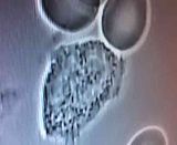 Here is a link to the German company that makes the blood laser equipment. www.webermedical.com/en/home/ |
|
|
|
Post by Baraka Obam on Aug 23, 2012 7:24:51 GMT -5
When I read that people were growing hair after the use of laser, I had a feeling it was destroying this hidden culprit.
I had ultra violet light that was actually put in a vein with a needle.
The thing I do have to wonder is, even if these light sources kill what is in the blood, it is just going to happen again because we have it living in our systems.
That is unless the youth of this monster only flows in the blood and after a time killing them keeps new infection from starting as the adult dies. I would assume.
|
|
bluesky
Junior Member
In memory of Dorothy
Posts: 63
|
Post by bluesky on Aug 23, 2012 10:59:14 GMT -5
I have no idea where it lives, other than what I can observe on my own body.
To be sure, I have scrutinized my live blood samples for any foreign organism, and other than bacteria, yeasts and a heart-stopping piece of (what turned out to be) dust on the slide, have seen nothing to attract paramedics, parasitologists or deities of any persuasion (Valhalla Starter Pac, exempted).
Baraka, if this is a nematomorph, as David thinks, it is likely in all the tissues, maybe or maybe not in the blood as well.
Can I hope that helping my immune system to get better will enable my immune system to get rid of it? Yes, I can hope, but that is not the same thing as saying that my immune system CAN or will be able to get rid of it.
I wonder if this (insert your own description of the cause of our malady) caused the damage to the RBC's that I saw in my blood samples? I wonder if it caused my immune system to not be working well. Because I have no live blood videos from before this mess began, I have no comparison, so cannot know.
I also wonder about the source of the bacteria in my samples. Is it carried by a possible organism that causes my illness; or is it the cause of my illness? No idea. And I am unlikely to get the bacteria identified because doctors here do not listen to, or take seriously, patients like me. Frankly, I suspect that they are terrified of us having relatives that are lawyers (LOL).
I only know that at times, for fleeting moments every once in a while, I feel like my old self again. I am choosing to do this documentation of this therapy here on LB as people here think outside the box -and I want people to see what I see from doing this. But urgently, too, in hopes that the treatment will lead to some relief from this, not just for me, but if it works, or even helps, it may help the hundreds of thousands of us who suffer with this. All very altruistic, yes, but selfish, too, because I desperately want to be well.
The practitioner I am working with thinks that this will rid my body of what is in my skin and scalp by rebuilding my immune system and blood system and correcting mitochondrial and cellular function. I hope that that is a correct viewpoint.
I do know of another person also doing a slightly different version of this UVB therapy, but who is also doing two other therapies simultaneously (which are not available to me at the moment). That other person is getting well slowly with time and continuing treatments of the 3 modalities. I would like to be doing the other two therapies at the same time as the blood laser therapy, but so far some of the components are missing. Perhaps in time that will change.
Baraka, I appreciate your comments, thoughts and musings. Some of them have brought a smile, many have echoed in my own head.
|
|
|
|
Post by Baraka Obam on Aug 23, 2012 13:27:27 GMT -5
I am also glad you are here laying out your trial for us.
The reasons I have these quetions to ask is because I had the intravenious Ultraviolet treatment in the vein.
They actually put this horse size needle into my arm and that needle had the light source inside it.
Along with this I got hydrogen peroxide, in the vein, vitamins, in the vein, serapeptase and 30 treatments of EDTA chelation.
There is one thing that happened with the EDTA I can say, I was growing these huge freakles on my shoulders, huge, they were light in color, when I got the EDTA chelation in the vein along with vitamins and minerals, the freckles vanished.
That is where all my questions came from.
I hope your treatment gives you lasting results that will be welcomed information .
|
|


