|
|
Post by kammy on Mar 22, 2010 16:43:54 GMT -5
Root colonization mycorhizes.com/Acolonisation.html"The objective of the observation and the estimation of mycorrhizal root colonization level is to determine the plant mycorrhizal status, to measure the mycorrhizal plant dependency toward arbuscular fungi and to compare colonization levels between treatments. The method proceed through extraction and surface cleaning of the plant root system followed by the bleaching of root tissue to clear them of cytoplasm and pigmentation and by the staining of root fungal structures. Once the roots are stained, their observation under the dissecting microscope or their mounting on slides for microscope closer observation allows to evaluate the level of root that are colonized, the type of fungal structures differentiated and to measure the abundance and the frequency of intraradical vesicles and arbuscules." What's a "intraradical vesicle"? journals.cambridge.org/action/displayAbstract?fromPage=online&aid=153811"Abstract Intraradical vesicles of Glomus intraradices were isolated, entrapped in alginate beads, and stored at 4 °C for periods from 2–74 months. The beads were used to inoculate leeks grown under standard conditions for 6 wk, then development of root colonization by G. intraradices was recorded. Colonization of leeks was high (mean >50% in length) and did not vary markedly until five years of storage. After six years of storage, the inoculum proved infective and viable." **Are they are putting alginated beads with fungi in them as standard practice for growing our root crops? Aren't alginated beads - nano? |
|
|
|
Post by kammy on Mar 22, 2010 17:06:24 GMT -5
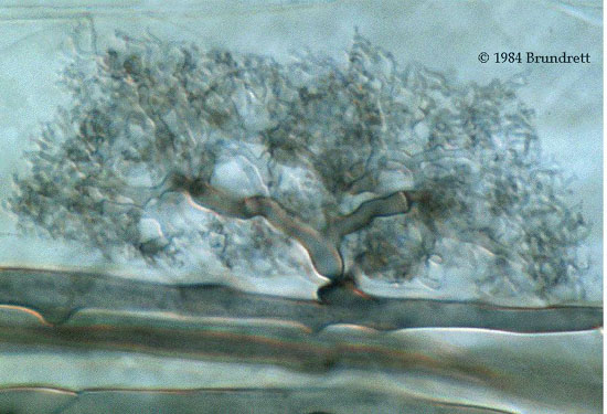
www.mycologia.org/cgi/content/full/97/6/1201/F5 "FIG. 5. Morphological features of G. proliferum/Gi. margarita (FIG. 5a–i) and G. intraradices/Gi. margarita (FIG. 5j–q) intraradical hyphae when colonizing common DC-2 roots. a–d. Plate 7, site A (closer to G. proliferum inoculum). a. Intercellular hyphae of G. proliferum (white arrow), together with arbuscules and some intraradical vesicles of this AMF (v) are clearly distinguishable. b. A closer view of G. proliferum arbuscules and vesicle (v). c and d. Two different views in the same picture in which a G. proliferum (white arrow) and a Gi. margarita (black arrow) entry points appear separated by a few micrometers. Note the different thickness of the extraradical hypha of each fungus. e–i. Plate 7, site B ( closer to Gi. margarita inoculum). e. Typical intraradical colonization by G. margarita, showing relatively large entry points and well defined arbuscules. f. A closer view of a Gi. margarita entry point (ep) and arbuscular colonization. g–i. Different views of a root site in which both G. proliferum and Gi. margarita intraradical colonization occur. g. General view of the site where auxiliary cells (aux) of Gi. margarita and a G. proliferum vesicle (v) coexist closely. Note the different hyphal thickness for both fungi (black and white arrows). h and i. Different focuses of a closer view of the site where intraradical hyphae of both fungi and a Gi. margarita branching event are clearly distinguishable. j–m. Plate 1, zone C (closer to G. intraradices inoculum). j. Typical G. intraradices arbuscular colonization. k. Paris-type colonization showing clear Gi. margarita arbusculate coils. l. Entry point (ep) and extensive colonization of a root by Gi. margarita. m. a closer view of the framed section where both arbusculate coils and typical coarse Gi. margarita extraradical hyphae are clearly distinguishable. n–q. Plate 1, zone D (closer to Gi. margarita inoculum). n and o. Extensive arbuscular colonization by Gi. margarita. Inset, a fan-like structure characteristic of Gi. margarita initial root recognition (arrow). p and q. Two sites where combined Gi. margarita (black arrows) and G. intraradices (solid black arrows) colonization is evident. aux, Gi. margarita auxiliary cells."  In "o." we see our little black 'carbon' balls... Gi. margarita.
|
|
|
|
Post by kammy on Mar 22, 2010 17:23:31 GMT -5
invam.caf.wvu.edu/fungi/taxonomy/Acaulosporaceae/Acaul_En.htmIs mode of spore formation enough to justify separation of species into two separate genera, Acaulospora and Entrophospora, in the family Acaulosporaceae? "The generic differences between Acaulospora and Entrophospora, much like those for Glomus and Sclerocystis, may not be as great as the current classification would suggest. The main property that separates the two genera is the position of the spore originating from the neck of the sporiferous saccule (a structure common to all members of the family Acaulosporaceae)." "Finally, the answer to the question posed in the title of this page is a conditional yes. From an evolutionary perspective, all organismal evidence indicates that Entrophospora is an artificial genus erected because of a very small developmental difference in position of spore formation that appears bigger than it really is when viewed statically as a morphological difference."
|
|
|
|
Post by kammy on Mar 22, 2010 17:40:30 GMT -5
Microscopy mycorhizes.com/Aobservation.html"The observation of colonized roots and of spores under the microscope is performed after mounting slides using or not histochemical reagents. Those reactives insure a better contrast of the studied material and facilitate the detection of the fine architecture of the fungal structures. Among others, the use of the Melzer’s reagent allows to better contrast some wpore walls and to detect the dextrinoVd nature of some of them. The Cotton Blue staining add to the better contrast of some spore structures. The use of phase contrast microscopy or of the differential interference contrast (Nomarski) can also refine the quality of the observation notably with surface ornamented spores." Ink and Vinegar www.ncbi.nlm.nih.gov/pmc/articles/PMC90956/"We developed a reliable, inexpensive, and simple method for staining arbuscular-mycorrhizal fungal colonizations in root tissues. Apart from applications in research, this nontoxic, high-quality staining method also could be of great utility in teaching exercises. After adequate clearing with KOH, an ink-vinegar solution successfully stained all fungal structures, rendering them clearly visible." |
|
|
|
Post by jeany on Mar 22, 2010 17:52:41 GMT -5
 I remember the 'morg-fungus-pix' you've shown us with the 'balls' sometimes on the side (Acaulospora) of the filament and some were within the filament (Entrophospora), bulb-looking. Jeany |
|
|
|
Post by jeany on Mar 22, 2010 19:14:55 GMT -5
CALCIUM ALGINATE BEADS AS A MATRIX FOR SLOW RELEASE OF PHEROMONES Calcium alginate beads are widely used for slow release of water soluble chemicals such as drugs, pesticides and fertilizers for about twenty years. The structure of the alginate, the physical and chemical properties of the beads, as well as their characteristics as a matrix for the slow release of water-soluble chemicals into an aqueous medium were intensively studied and are well known. In the present work we investigated the parameters that govern the slow release characteristics of water-insoluble materials, as emulsion droplets, from a matrix of calcium alginate into the atmosphere, using gelatin as the surface-active agent in the emulsification stage.
The motivation behind the research is the possibility to use the formulation of calcium alginate/gelatin/water-insoluble-material as a matrix for the slow release of biologically active materials, such as pheromones, in integrated pest management. * pheromones? no wonder we attract bugs! www.weizmann.ac.il/ICS/chemistry66/142Yosha.html |
|
|
|
Post by jeany on Mar 22, 2010 19:19:06 GMT -5
**Are they are putting alginated beads with fungi in them as standard practice for growing our root crops? Aren't alginated beads - nano? Molecular biologists are using this common technique. Dynabeads are magnetic and have polymer shell! Characteristics of Dynabeads®Dynabeads® are superparamagnetic particles, meaning that the beads exhibit magnetic properties in a magnetic field with no residual magnetism once removed Magentic bead separation is gentle and no columns or centrifugations are necessary Spherical shape and defined surface chemistry minimize chemical agglutination and non-specific binding True uniformity (CV<3%) of size, shape and surface area provides optimal accessibility and reaction kinetics, for rapid and efficient binding Unique batch-to-batch reproducibility (typically within 5%) secures reproducible, quality-results The polymer shell on each bead protects your target from toxic exposure to iron Specific characteristics of the many available bead types offer magnetic separation of a variety of targets Dynabeads® Types and UsesDynabeads® come in several sizes and with different surface functionalities, for use in a wide variety of applications.  Some beads are pre-coupled with a biomolecule (ligand). The ligand can be an antibody, protein or antigen, DNA/RNA probe or any other molecule with an affinity for the desired target. Ready-to-use Dynabeads products and kits are available for many applications. For assay developments that require flexibility, a range of Dynabeads products with specific characteristics are available. How to use Dynabeads®The Dynabeads magnetic separation protocol consists of three simple steps: Bind When added to a sample, Dynabeads® bind to the desired target (cells, pathogenic microorganisms, nucleic acids, peptide, protein or protein complex etc). This interaction relies on the specific affinity of the ligand on the surface of the beads. Wash The beads respond to a magnetic field, allowing bound material to be rapidly and efficiently separated from the rest of the sample. Unbound material is simply removed by aspiration, and the bead-bound target washed by the use of the magnet. Elute The bead-bound target is released in a suitable volume for use in downstream applications. Alternatively, the bead-bound target can be used directly while still attached to the beads. No centrifugation or columns are required. The Dynabeads magnetic separation protocol is scalable and very automation-friendly. www.invitrogen.com/site/us/en/home/brands/Dynal/dynabeads_technology.html |
|
|
|
Post by kammy on Mar 22, 2010 22:29:04 GMT -5
I want to thank Tam Tam and Skytroll for helping me get to yesterday's discovery of the Morgellons fungi and the process behind it.
|
|
|
|
Post by skytroll on Mar 23, 2010 1:18:01 GMT -5
clathrin coated vesicles
Clathrin is a protein which plays a major role in the formation of vesicles. ....isolated and named by Barbara Pearse in 1975. It forms a triskelion shape which is composed of three clathrin heavy chains and three light chains. When the triskelion interact they form a polyhedral lattice which surrounds the vesicle.
from wikepedia, my linker does not work.
skytroll
|
|
|
|
Post by kammy on Mar 23, 2010 3:04:51 GMT -5
Hi Sky, we're glad you're here! I thought that Clathrin was a naturally occurring protein in the body? en.wikipedia.org/wiki/ClathrinI see hexagonal patterns everywhere... see how the molecule is made up of hexagons to form the vesicle? We can't see this in the photos above, not enough magnification.  |
|
|
|
Post by kammy on Mar 23, 2010 5:48:42 GMT -5
|
|
|
|
Post by kammy on Mar 23, 2010 7:21:56 GMT -5
Freaky is about to see her doctor who is Infectious Diseases, I believe? I'm in the process of finding out her specialty and Freaky asked me which sites she should refer the doctor to? I think the morgellons.org/case_definition.htm site has the best, compact information for a doctor? Freaky, you can cut and paste this below or if folks have a suggested revision?... Dear Dr. Salvato, Morgellons Disease is poorly understood at this time, one of our best resources is the morgellons.org/case_definition.htm page written by a Medical Board to inform doctors of the Case History studies of several Morgellons patients that were evaluated. In this researcher's opinion, it appears that Morgellons disease is strongly related to a systemic fungal/bacterial infection related to the Mycorrhizal genus. This fungus is not being found in traditional lab testing because these fungi are used in fertilizers and food fermentation processing and are generally regarded as safe. The Morgellons patient needs a referral to a Mycologist. |
|
|
|
Post by kammy on Mar 23, 2010 7:26:01 GMT -5
Of course, a form letter to the Mycologist would read like a whole different story!...
|
|
|
|
Post by kammy on Mar 23, 2010 10:05:38 GMT -5
Does anyone have contact with Becky McClain that could email her a link to a thread on Morgellons to get her to look at it? Would you PM me, please, if you do?
|
|
|
|
Post by rhorn2006 on Mar 23, 2010 10:37:48 GMT -5
Kammy.... I ran a search and the only contact information I could find "related to Becky McClain" is through a site that hosts some of her documents "The Center for Public Awareness in Bioethics (CPAB)" Contact: cpab.info/contactus.aspxMaybe they will get word to her for you.. _________________________________________________________________________ Where I got this address info.. www.theprogressivemind.info/?p=18194 There are other addresses in the story that might help you contact her... Story title: The Nightmare At Pfizer - Injured Biotech Worker Becky McClain Speaks at 2009 SF Workers Memorial Day. <last paragraph> In light of the new emerging swine flu epidemic in Mexico, biotech workers rights and public health and safety are the first line of defense against accidental, negligent or intentional release of infectious agents that could cause new emerging disease. Becky also talk about the case of biotech worker Dr. Jeannette Adu-Bobie and how she had to fight for healthcare after being infected at her laboratory in New Zealand. You can find more out about McClain’s work at www.workersmemorialday.org/documents/McClain.htm www.cpab.info director@cpab.info The San Francisco Workers Memorial Day event on April 28, 2009 where McClain spoke was sponsored by California Coalition For Workers Memorial Day and was held at ILWU Local 34 in San Francisco. www.workersmemorialday.org Video Produced by Labor Video Project P.O. Box 720027 San Francisco, CA 94172 www.laborvideo.orgThe Nightmare At Pfizer - Injured Biotech Worker Becky McClain Speaks at 2009 SF Workers Memorial Day. |
|
|
|
Post by kammy on Mar 23, 2010 11:40:54 GMT -5
TY, Ron!
I think a couple of the gals know her and talk to her once in a while in emails? I know it's a bad time for her with her court case coming up...? and all...
|
|
|
|
Post by kammy on Mar 23, 2010 12:04:17 GMT -5
"The Cones" We know the nature of some of our lesions has been referred to as like a 'cone' that is embedded in the skin. That some seem to be funnel or tunnel-like and with a root system, we can't easily remove them, in fact - they are almost impossible to remove. I'm seeing where this is a particular trait of the mycorrhiza fungi called Arbuscular mycorrhiza or "AM fungi". en.wikipedia.org/wiki/Arbuscular_mycorrhiza"An arbuscular mycorrhiza (plural mycorrhizae or mycorrhizas) is a type of mycorrhiza in which the fungus penetrates the cortical cells of the roots of a vascular plant. Arbuscular mycorrhizae (AMs) are characterized by the formation of unique structures such as arbuscules and vesicles by fungi of the phylum Glomeromycota (AM fungi). AM fungi help plants to capture nutrients such as phosphorus and micronutrients from the soil. It is believed that the development of the arbuscular mycorrhizal symbiosis played a crucial role in the initial colonisation of land by plants and in the evolution of the vascular plants.[1] It has been said that it is quicker to list the plants that do not form mycorrhizae than those that do.[2] This symbiosis is a highly evolved mutualistic relationship found between fungi and plants, the most prevalent plant symbiosis known,[3] and AM is found in 80% of vascular plant families of today.[4]" Here's what an "AM" stock photo looks like: I'm saying the 'cones' resemble this nodule above from which the branches are forming down into and under our skin. The only problem is... this "AM" is only supposed to happen in plants. www.soilhealth.com/fungi/#1"Arbuscular mycorrhizas are called this because they have developed a specific structure called an "arbuscule". Although some kinds of soil fungi can be associated with plant and animal disease, the fungi that form arbuscular mycorrhizas belong to a group of soil fungi that can be very beneficial. The name "mycorrhiza" means "fungus root" and this is derived from the close association of the fungi with plant roots - in fact, arbuscular mycorrhizal fungi cannot complete their life cycle unless they are connected to plant roots. Generally, it is not possible to grow these fungi without the support of the plant. The reasons for this are still largely unknown." |
|
|
|
Post by kammy on Mar 23, 2010 12:14:23 GMT -5
Close To The Source Well... what happened? My ear is the leaf on a plant? It's riddled with the species within this group that causes the formation of the unique structures called "arbuscules". If we look at the patents we can see where the scientists 'souped up' this arbuscules formation process. When the particles sprouted out of my head that landed on my body and ear... they were highly programmed to burrow deep into my skin, to create the arbuscules with a multi-hyphaed, deeply embedded root system from it. |
|
jlk
Full Member

Posts: 146
|
Post by jlk on Mar 23, 2010 18:19:34 GMT -5
Kammy, and all you smart people - thanks so much for doing the work that should have been done by those entrusted with "protecting our health". I was writing the book that is now available for free at judithnd.com and I will be updating it with a link to these blog pages because you have all nailed it. I don't think it will be anytime soon, even if you guys did all the work for them, that the powers that be will do anything about this. Also, since I own the e-book if any of you have additional information you want me to add to it as an afterward 1- 2- 3- or put some links up on my site I welcome your contributions. Further, I have contacted Larry King, Oprah, The View, CNN, Mike Moore and there will be recognition as it will get into somebody's hands that is going to bring this to the forefront again. I was on Dr Phil long ago and I will get on somewhere again. You will not be doing all this work for nothing - please do not feel that way. You are all part of the tapestry that forms the whole picture every thread and weave makes it come together. Those who have been on this site for a long time know I was on here awhile back - my mom got cancer for the third time at the age of 85 last year and I stopped most of my online blogs, reading posting etc... she is now 86 and though the cancer is not back yet she is slowly going down mentally so I have responsibility at home but I will do all I can to push this book and thus bring greater awareness to all of your efforts. That is why I did this. Any of you can contact me at judithnd(capital 2)judithnd.com with additional information related to this posting - topic. I think the big corporate guys will keep this under cover as long as they can but I am going to do all I can to expose this mess to the world. We have been had. This stuff has destroyed the entire ecosystem and it is only a matter of time before hundreds of thousands more will be wondering "why are these sores not healing?" That is why I am determined to get this book out there - I want them to know the truth. To get better, if you read the book, you will learn this: MOST IMPORTANT first steps DO NOT GO AFTER TREATING THE SKIN BEFORE YOU ARE ATTACKING IT INTERNALLY you will just drive it deeper if you go after it externally. I had the gel form lumps all over my body and as I continued to study I learned that I could dissolve biofilms, (and this gel) with special enzymes. Once I got on it (you'll love this) my sores got way worse, I had blisters form on my fingers and organisms came out like crazy - as did the fibers and capsids - I got very fatigued. This is what it takes to get rid of this crap. The enzymes broke open the "safe houses" of the organisms and they came out of me like crazy. It is not fun but that is what must happen. Lyme is exactly the same - you have to get rid of the biofilms so that you can get at the various organims. At the same time I was then using the professional detoxification products offer by DNR Inc. It is explained in the back of the book. No one gets better until they destroy the biofilm - I tell you how - then you kill the various pathogens and get rid of the toxins - then you can enjoy a better life. Cured??? If you can do this within 2-3 years of exposure I would say yes - but if you have had it as long as I have (12-15 yrs) it is deep in all my tissues, spine, bones, lungs... so I am still working on it but I do have a better quality of life than I had before doing this. For external skin and household the best product is Best Yet from CedarCide - red cedar essential oil is in it. Great stuff for PETS - clears them up fast! Biofilm product can be used internally for pets too. Then, today as I walked around my yard I found huge piles of fungus that had been under a snow pile and I know it is this stuff. So this summer when I mow the grass I will breathe that all in again. I am not sure there will be any escape if one breathes and uses water. It is in the well water at my place. How do I know this: I have seen hundreds of times the instant self-assembly of the fibers in my sink or tub and so I thought they were coming from my skin cells, as I shed this stuff. But, I also have a cottage that I rent out to others and had not been in it for a long time and before the guy moved in it had been completely remodeled - new sinks, new walls mostly gutted and redone - well to make a long story short - I had to clean it up when he moved out and I had not been in it for even 1 minute before I began cleaning the bathroom sink - what formed instantly - you guessed it - the fibers - they are from the water (the same well water is in my house as the cottage) - they are not only forming from me - they are forming from the water. So we are screwed! This crap has gotten into the ground water and nobody is aware of what is going on. Read the book - make sure to share it - get it into newsletters - we have to warn the public what a nightmare this is. Thanks everyone - you all rock! Judy |
|
|
|
Post by skytroll on Mar 23, 2010 23:55:41 GMT -5
There is much more to this then just the fungi, Kammy. How does this get into the human body? What is the delivery system? Has this fungus been found on the human body? How does it relate to chlamydia? What is the submicron particle? How do erthrocytes fit in here? I think we have to take this further yet, bannany just found from her report Feather particles, which tam tam mentioned at one time, spiders and webs found around home smuts and myxomycetes, agro is rhizobiums. going to root of plant, and seed. plant parts textile parts gell shards. Quorum sensing has to be in the carbon ball itself, made most likely of more than carbon magnetic like fullerene used in buckyballs, remember these are very small. and they can carry dna. Metalloproteins. Yet they can react to both candida yeasts teliospores from smut and rust. candida is like conidia and basidiomycetes. There is the heat shock proteins that are making certain genetic types very ill. The new artficial cell, includes a natural clathrin coat, or an inorganic one made of iron, fullerenes, magnetic, and that is in the carbon buckyball. forms the cage also in nanotubes. This was all part of a DOE project where microbes were created to eat up stuff in the environment. Part of the aerosol operations. What we have cannot be cultivated because of the Archeae in the construction that involves thermomycota, rhodopsin, and other thermophiles. This crosses the bacteria, archaea, and eukaryotics which involve man and yeast and fungi is closest to genes in man. I think we are looking at yeast, this is what forms the chlamydia forms. This yeast is close to smut and rust diseases. What are the spores seed that makes up this chlamydia? It is related to candida yeast, the 6 that form chlamydia and the yeast artificial chromosome. These have the power to change our histones adding a 5th histone. or more. Smut and rust teliospores are very close to the forms. ====================== Origins and inheritance of chromosome-length polymorphisms in the ... by DA Gaudet - 1998 - Cited by 8 - Related articles (teliospore line 1279). Polymorphisms for chromosome IV were observed in the 1600–1900-kb ...... insertions within telomeres contribute to chromosome-size ... www.springerlink.com/index/TDY4VYFR0FJAEU2B.pdfoff google compare telomeres in human body to teliospores in uridinales. Hang in there, we are not there yet. we need proof. specimens, not photos. We need to know how and why the genetists decided to reconstruct the cytoskeleton system. Homologenous genes substituting for human genes. ................ purposely..................... skytroll |
|